Heußer Thorsten, Mann Philipp, Rank Christopher M, Schäfer Martin, Dimitrakopoulou-Strauss Antonia, Schlemmer Heinz-Peter, Hadaschik Boris A, Kopka Klaus, Bachert Peter, Kachelrieß Marc, Freitag Martin T
Medical Physics in Radiology, German Cancer Research Center (DKFZ), Heidelberg, Germany.
Applied Medical Radiation Physics, German Cancer Research Center (DKFZ), Heidelberg, Germany.
PLoS One. 2017 Aug 17;12(8):e0183329. doi: 10.1371/journal.pone.0183329. eCollection 2017.
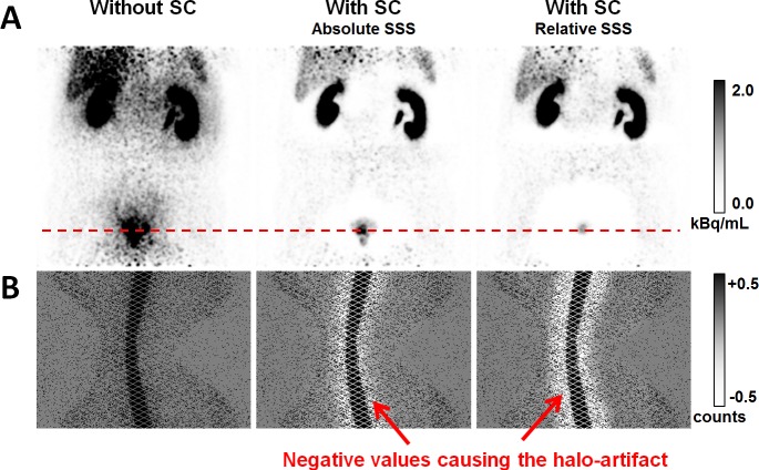
Combined positron emission tomography (PET) and magnetic resonance imaging (MRI) targeting the prostate-specific membrane antigen (PSMA) with a 68Ga-labelled PSMA-analog (68Ga-PSMA-11) is discussed as a promising diagnostic method for patients with suspicion or history of prostate cancer. One potential drawback of this method are severe photopenic (halo-) artifacts surrounding the bladder and the kidneys in the scatter-corrected PET images, which have been reported to occur frequently in clinical practice. The goal of this work was to investigate the occurrence and impact of these artifacts and, secondly, to evaluate variants of the standard scatter correction method with regard to halo-artifact suppression.
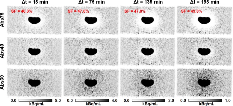
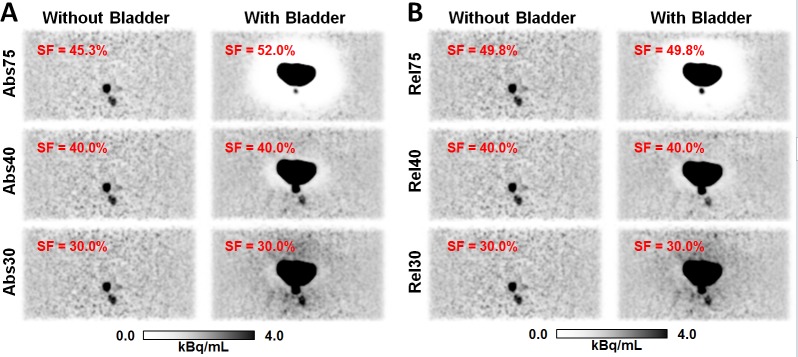
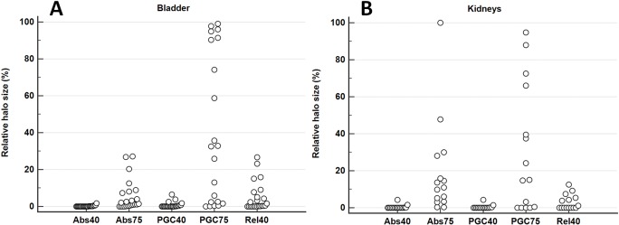
Experiments using a dedicated pelvis phantom were conducted to investigate whether the halo-artifact is modality-, tracer-, and/or concentration-dependent. Furthermore, 31 patients with history of prostate cancer were selected from an ongoing 68Ga-PSMA-11-PET/MRI study. For each patient, PET raw data were reconstructed employing six different variants of PET scatter correction: absolute scatter scaling, relative scatter scaling, and relative scatter scaling combined with prompt gamma correction, each of which was combined with a maximum scatter fraction (MaxSF) of MaxSF = 75% or MaxSF = 40%. Evaluation of the reconstructed images with regard to halo-artifact suppression was performed both quantitatively using statistical analysis and qualitatively by two independent readers.
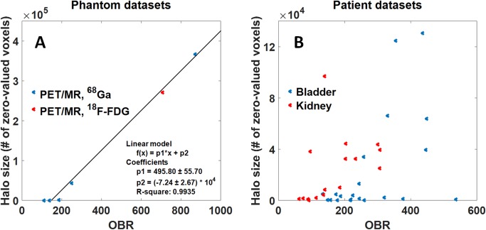
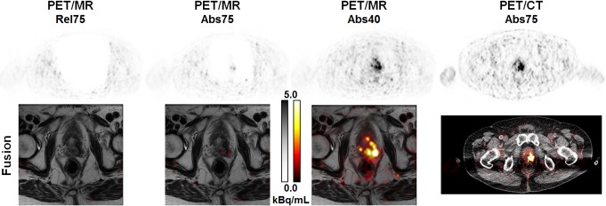
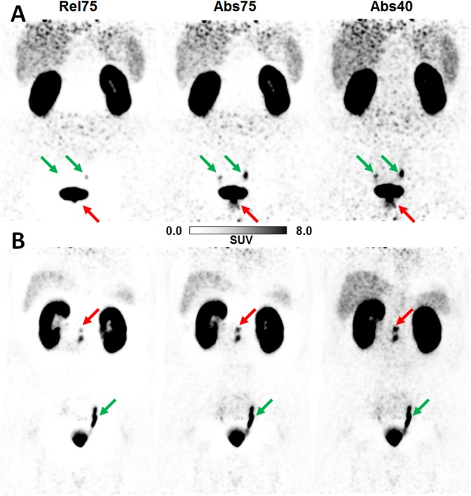
The phantom experiments did not reveal any modality-dependency (PET/MRI vs. PET/CT) or tracer-dependency (68Ga vs. 18F-FDG). Patient- and phantom-based data indicated that halo-artifacts derive from high organ-to-background activity ratios (OBR) between bladder/kidneys and surrounding soft tissue, with a positive correlation between OBR and halo size. Comparing different variants of scatter correction, reducing the maximum scatter fraction from the default value MaxSF = 75% to MaxSF = 40% was found to efficiently suppress halo-artifacts in both phantom and patient data. In 1 of 31 patients, reducing the maximum scatter fraction provided new PET-based information changing the patient's diagnosis.
Halo-artifacts are particularly observed for 68Ga-PSMA-11-PET/MRI due to 1) the biodistribution of the PSMA-11-tracer resulting in large OBRs for bladder and kidneys and 2) inaccurate scatter correction methods currently used in clinical routine, which tend to overestimate the scatter contribution. If not compensated for, 68Ga-PSMA-11 uptake pathologies may be masked by halo-artifacts leading to false-negative diagnoses. Reducing the maximum scatter fraction was found to efficiently suppress halo-artifacts.
正电子发射断层扫描(PET)与磁共振成像(MRI)相结合,使用68Ga标记的前列腺特异性膜抗原(PSMA)类似物(68Ga-PSMA-11)靶向前列腺,被认为是一种对疑似患有前列腺癌或有前列腺癌病史的患者很有前景的诊断方法。这种方法的一个潜在缺点是,在散射校正的PET图像中,膀胱和肾脏周围会出现严重的放射性缺损(晕圈)伪影,据报道在临床实践中经常出现。这项工作的目的是研究这些伪影的发生情况及其影响,其次,评估标准散射校正方法在抑制晕圈伪影方面的变体。
使用专用的骨盆模型进行实验,以研究晕圈伪影是否与模态、示踪剂和/或浓度有关。此外,从正在进行的68Ga-PSMA-11-PET/MRI研究中挑选出31例有前列腺癌病史的患者。对于每位患者,使用六种不同的PET散射校正变体重建PET原始数据:绝对散射缩放、相对散射缩放以及结合了瞬发伽马校正的相对散射缩放,每种方法都与最大散射分数(MaxSF)为75%或40%相结合。通过统计分析对重建图像进行晕圈伪影抑制的定量评估,并由两名独立的阅片者进行定性评估。
模型实验未发现任何模态依赖性(PET/MRI与PET/CT)或示踪剂依赖性(68Ga与18F-FDG)。基于患者和模型的数据表明,晕圈伪影源自膀胱/肾脏与周围软组织之间较高的器官与本底活性比(OBR),且OBR与晕圈大小呈正相关。比较不同的散射校正变体,发现将最大散射分数从默认值MaxSF = 75%降低到MaxSF = 40%可有效抑制模型和患者数据中的晕圈伪影。在31例患者中的1例中,降低最大散射分数提供了基于PET的新信息,改变了患者的诊断。
对于68Ga-PSMA-11-PET/MRI,特别容易观察到晕圈伪影,原因如下:1)PSMA-11示踪剂的生物分布导致膀胱和肾脏的OBR较大;2)临床常规中目前使用的散射校正方法不准确,往往会高估散射贡献。如果不进行补偿,68Ga-PSMA-11摄取异常可能会被晕圈伪影掩盖,导致假阴性诊断。发现降低最大散射分数可有效抑制晕圈伪影。