• 文献检索
  • 文档翻译
  • 深度研究
  • 学术资讯
  • Suppr Zotero 插件Zotero 插件
  • 邀请有礼
  • 套餐&价格
  • 历史记录
应用&插件
Suppr Zotero 插件Zotero 插件浏览器插件Mac 客户端Windows 客户端微信小程序
定价
高级版会员购买积分包购买API积分包
服务
文献检索文档翻译深度研究API 文档MCP 服务
关于我们
关于 Suppr公司介绍联系我们用户协议隐私条款
关注我们

Suppr 超能文献

核心技术专利:CN118964589B侵权必究
粤ICP备2023148730 号-1Suppr @ 2026

文献检索

告别复杂PubMed语法,用中文像聊天一样搜索,搜遍4000万医学文献。AI智能推荐,让科研检索更轻松。

立即免费搜索

文件翻译

保留排版,准确专业,支持PDF/Word/PPT等文件格式,支持 12+语言互译。

免费翻译文档

深度研究

AI帮你快速写综述,25分钟生成高质量综述,智能提取关键信息,辅助科研写作。

立即免费体验

多层螺旋计算机断层扫描与传统冠状动脉造影术在检测支架内再狭窄方面的价值比较

The Value of Multi-slice Computed Tomography Compared to Conventional Coronary Angiography for Detecting In-stent Restenosis.

作者信息

Karami Mehdi, Sajjadieh Amirreza, Moradi Ahmad, Taleb Akbar, Brumand Sareh

机构信息

Department of Radiology, Isfahan University of Medical Sciences, Isfahan, Iran.

出版信息

Adv Biomed Res. 2017 Jul 28;6:92. doi: 10.4103/2277-9175.211796. eCollection 2017.

DOI:10.4103/2277-9175.211796
PMID:28828343
原文链接:https://pmc.ncbi.nlm.nih.gov/articles/PMC5549549/
Abstract

BACKGROUND

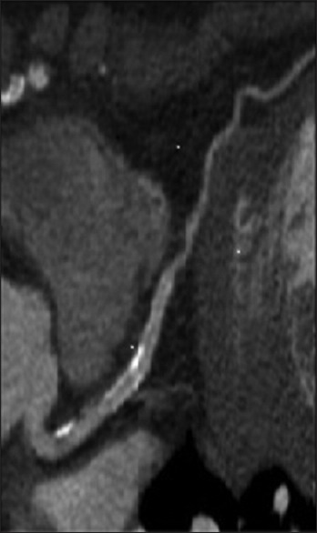
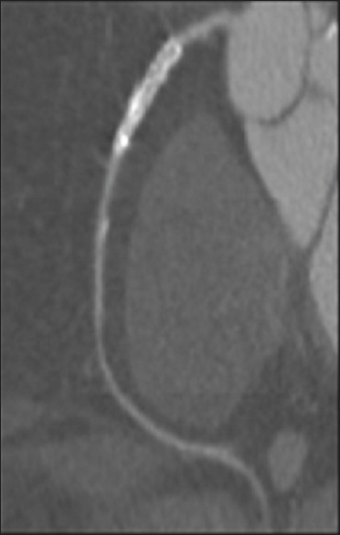
Diagnostic value of multi-slice computed tomography (MSCT) for detecting in-stent restenosis in comparison with conventional coronary angiography remained uncertain. The present study aimed to determine the value of MSCT for detecting in-stent restenosis.

MATERIALS AND METHODS

This historical cohort study was included 226 patients with the history of percutaneous coronary intervention from 2000 to 2014 that referred to MSCT Unit at Alzahra Heart Center in Isfahan. The subjects were followed-up by telephone with regard to performing coronary angiography up to 3 months after MSCT and their status about cardiac events.

RESULTS

Among all participants, 63 stents (27.9%) underwent coronary angiography up to 3 months after MSCT that 2 stents in left circumflex artery (LCX) assessment, 2 in left anterior descending (LAD) assessments and none in right coronary artery (RCA) assessment were uninterpretable. Sensitivity, specificity, positive predictive value (PPV), negative predictive value (NPV), and accuracy of MSCT was 92.9%, 66.6%. 92.9%, 66.6%, and 88.2%, respectively for detection of occlusion in LCX stents, 100%, 100%, 100%, 100%, and 100%, respectively for detection of occlusion in LAD stents, and 80.0%, 0.0%, 80.0%, 0.0%, and 66.7%, respectively for detection of occlusion in RCA stents. Overall, MSCT had sensitivity of 93.8%, specificity of 70.0%, PPV of 93.8%, NPV of 70.0%, and accuracy 89.7% for detection of coronary stent restenosis.

CONCLUSION

MSCT has high diagnostic value for detecting in-stent restenosis. Diagnostic accuracy of MSCT for detecting stent restenosis is considerably different between the coronary arteries with the highest diagnostic values for LAD and the lowest diagnostic values for RCA.

摘要

背景

与传统冠状动脉造影相比,多层螺旋计算机断层扫描(MSCT)检测支架内再狭窄的诊断价值仍不确定。本研究旨在确定MSCT检测支架内再狭窄的价值。

材料与方法

这项历史性队列研究纳入了2000年至2014年期间在伊斯法罕的阿尔扎赫拉心脏中心MSCT科接受经皮冠状动脉介入治疗的226例患者。通过电话随访受试者,了解其在MSCT后3个月内进行冠状动脉造影的情况以及心脏事件状态。

结果

在所有参与者中,63个支架(27.9%)在MSCT后3个月内接受了冠状动脉造影,其中左旋支动脉(LCX)评估中有2个支架、左前降支(LAD)评估中有2个支架、右冠状动脉(RCA)评估中无支架无法解读。MSCT检测LCX支架闭塞的敏感性、特异性、阳性预测值(PPV)、阴性预测值(NPV)和准确性分别为92.9%、66.6%、92.9%、66.6%和88.2%;检测LAD支架闭塞的敏感性、特异性、PPV、NPV和准确性分别为100%、100%、100%、100%和100%;检测RCA支架闭塞的敏感性、特异性、PPV、NPV和准确性分别为80.0%、0.0%、80.0%、0.0%和66.7%。总体而言,MSCT检测冠状动脉支架再狭窄的敏感性为93.8%,特异性为70.0%,PPV为93.8%,NPV为70.0%,准确性为89.7%。

结论

MSCT检测支架内再狭窄具有较高的诊断价值。MSCT检测支架再狭窄的诊断准确性在LAD诊断价值最高和RCA诊断价值最低的冠状动脉之间存在显著差异。

https://cdn.ncbi.nlm.nih.gov/pmc/blobs/127d/5549549/26b6fcff5a78/ABR-6-92-g002.jpg
https://cdn.ncbi.nlm.nih.gov/pmc/blobs/127d/5549549/a7c32ca6947c/ABR-6-92-g001.jpg
https://cdn.ncbi.nlm.nih.gov/pmc/blobs/127d/5549549/26b6fcff5a78/ABR-6-92-g002.jpg
https://cdn.ncbi.nlm.nih.gov/pmc/blobs/127d/5549549/a7c32ca6947c/ABR-6-92-g001.jpg
https://cdn.ncbi.nlm.nih.gov/pmc/blobs/127d/5549549/26b6fcff5a78/ABR-6-92-g002.jpg

相似文献

1
The Value of Multi-slice Computed Tomography Compared to Conventional Coronary Angiography for Detecting In-stent Restenosis.多层螺旋计算机断层扫描与传统冠状动脉造影术在检测支架内再狭窄方面的价值比较
Adv Biomed Res. 2017 Jul 28;6:92. doi: 10.4103/2277-9175.211796. eCollection 2017.
2
Usefulness of multislice computed tomography to assess patency of coronary artery stents versus conventional coronary angiography.多层螺旋计算机断层扫描与传统冠状动脉造影术评估冠状动脉支架通畅性的效用比较
J Cardiovasc Med (Hagerstown). 2008 May;9(5):485-92. doi: 10.2459/JCM.0b013e3282f2c9e3.
3
Usefulness of 64-slice multislice computed tomography coronary angiography to assess in-stent restenosis.64层多层螺旋CT冠状动脉造影评估支架内再狭窄的效用。
J Am Coll Cardiol. 2007 Jun 5;49(22):2204-10. doi: 10.1016/j.jacc.2007.02.045. Epub 2007 May 23.
4
Impairment factors for evaluating the patency of drug-eluting stents and bare metal stents in coronary arteries by 64-slice computed tomography versus conventional coronary angiography.64层计算机断层扫描与传统冠状动脉造影术评估药物洗脱支架和裸金属支架在冠状动脉中通畅性的损伤因素
Int J Cardiol. 2008 Nov 28;130(3):349-56. doi: 10.1016/j.ijcard.2007.08.104. Epub 2008 Jan 3.
5
Comparative analysis between 64- and 320-slice spiral computed tomography in the display of coronary artery stents and diagnosis of in-stent restenosis.64层与320层螺旋计算机断层扫描在冠状动脉支架显示及支架内再狭窄诊断中的对比分析
Exp Ther Med. 2015 Nov;10(5):1871-1876. doi: 10.3892/etm.2015.2768. Epub 2015 Sep 23.
6
Multislice spiral computed tomography for the evaluation of stent patency after left main coronary artery stenting: a comparison with conventional coronary angiography and intravascular ultrasound.多层螺旋计算机断层扫描用于评估左主干冠状动脉支架置入术后支架通畅情况:与传统冠状动脉造影和血管内超声的比较
Circulation. 2006 Aug 15;114(7):645-53. doi: 10.1161/CIRCULATIONAHA.105.608950. Epub 2006 Aug 7.
7
Accuracy of multislice computed tomography coronary angiography: a pooled estimate.多层螺旋计算机断层扫描冠状动脉造影的准确性:一项汇总估计。
Cardiol Rev. 2008 Jul-Aug;16(4):189-96. doi: 10.1097/CRD.0b013e318164d02a.
8
Heart imaging: the accuracy of the 64-MSCT in the detection of coronary artery disease.心脏成像:64层螺旋CT检测冠状动脉疾病的准确性
Eur Rev Med Pharmacol Sci. 2009 May-Jun;13(3):163-71.
9
Systematic review of the clinical effectiveness and cost-effectiveness of 64-slice or higher computed tomography angiography as an alternative to invasive coronary angiography in the investigation of coronary artery disease.64层及以上计算机断层扫描血管造影术作为冠状动脉疾病检查中侵入性冠状动脉造影替代方法的临床有效性和成本效益的系统评价。
Health Technol Assess. 2008 May;12(17):iii-iv, ix-143. doi: 10.3310/hta12170.
10
Evaluation of coronary artery in-stent restenosis by 64-section computed tomography: factors affecting assessment and accurate diagnosis.64 层 CT 评价冠状动脉支架内再狭窄:影响评估和准确诊断的因素。
J Thorac Imaging. 2010 Feb;25(1):57-63. doi: 10.1097/RTI.0b013e3181b5d813.

本文引用的文献

1
New quantitative method to diagnose coronary in-stent restenosis by 64-multislice computed tomography.通过64层螺旋计算机断层扫描诊断冠状动脉支架内再狭窄的新定量方法。
J Cardiol. 2015 Jan;65(1):57-62. doi: 10.1016/j.jjcc.2014.03.013. Epub 2014 May 18.
2
[Predictors of in-stent restenosis in coronary heart disease patients complicating with diabetes mellitus within 2 years after drug-eluting stents implantation].[药物洗脱支架植入术后2年内合并糖尿病的冠心病患者支架内再狭窄的预测因素]
Zhonghua Xin Xue Guan Bing Za Zhi. 2014 Jan;42(1):14-8.
3
Incidence and predictors of restenosis after coronary stenting in 10 004 patients with surveillance angiography.
10004 例接受监测血管造影的患者冠状动脉支架置入术后再狭窄的发生率和预测因素。
Heart. 2014 Jan;100(2):153-9. doi: 10.1136/heartjnl-2013-304933. Epub 2013 Nov 22.
4
Computed tomography angiography and myocardial computed tomography perfusion in patients with coronary stents: prospective intraindividual comparison with conventional coronary angiography.冠状动脉支架置入术后患者的 CT 血管造影和心肌 CT 灌注:与常规冠状动脉造影的前瞻性个体化比较。
J Am Coll Cardiol. 2013 Oct 15;62(16):1476-85. doi: 10.1016/j.jacc.2013.03.088. Epub 2013 Jun 19.
5
Diagnostic performance of 64-row coronary CT angiography in detecting significant stenosis as compared with conventional invasive coronary angiography.与传统有创冠状动脉造影术相比,64排冠状动脉CT血管造影术检测显著狭窄的诊断性能。
ARYA Atheroscler. 2013 Mar;9(2):157-63.
6
Coronary in-stent restenosis: assessment with CT coronary angiography.冠状动脉支架内再狭窄:CT 冠状动脉成像评估。
Radiology. 2012 Nov;265(2):410-7. doi: 10.1148/radiol.12112363. Epub 2012 Sep 10.
7
Incidence of definite stent thrombosis or in-stent restenosis after drug-eluting stent implantation for treatment of coronary in-stent restenosis: from Western Denmark Heart Registry.药物洗脱支架置入治疗冠状动脉支架内再狭窄后明确的支架血栓形成或支架内再狭窄发生率:来自丹麦西部心脏注册研究。
Catheter Cardiovasc Interv. 2013 Feb;81(2):260-5. doi: 10.1002/ccd.24398. Epub 2012 Nov 8.
8
Role of Stress Myocardial Scintigraphy in the Evaluation of Incompletely Revascularized Post-PCI Patients.应激心肌显像在PCI术后血运重建不完全患者评估中的作用
Int J Mol Imaging. 2011;2011:180936. doi: 10.1155/2011/180936. Epub 2011 Sep 21.
9
[Value of detecting in-stent restenosis by dual source coronary computed tomography coronary angiography].[双源冠状动脉计算机断层扫描冠状动脉造影检测支架内再狭窄的价值]
Zhonghua Xin Xue Guan Bing Za Zhi. 2011 Jan;39(1):49-52.
10
The diagnostic accuracy of 256-row computed tomographic angiography compared with invasive coronary angiography in patients with suspected coronary artery disease.256 排计算机断层血管造影与有创冠状动脉造影诊断疑似冠心病的准确性比较。
Eur Heart J. 2010 Aug;31(15):1916-23. doi: 10.1093/eurheartj/ehq072. Epub 2010 Mar 16.