Vidorreta Marta, Wang Ze, Chang Yulin V, Wolk David A, Fernández-Seara María A, Detre John A
Department of Neurology, University of Pennsylvania, Philadelphia, Pennsylvania, United States of America.
Department of Radiology, University of Pennsylvania, Philadelphia, Pennsylvania, United States of America.
PLoS One. 2017 Aug 24;12(8):e0183762. doi: 10.1371/journal.pone.0183762. eCollection 2017.
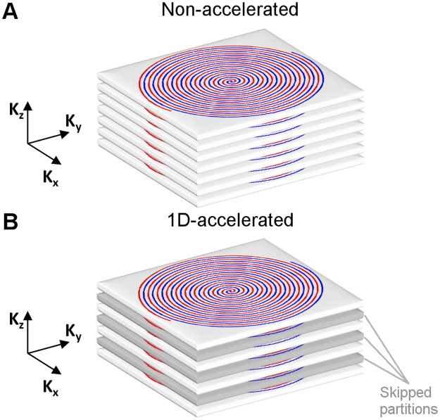
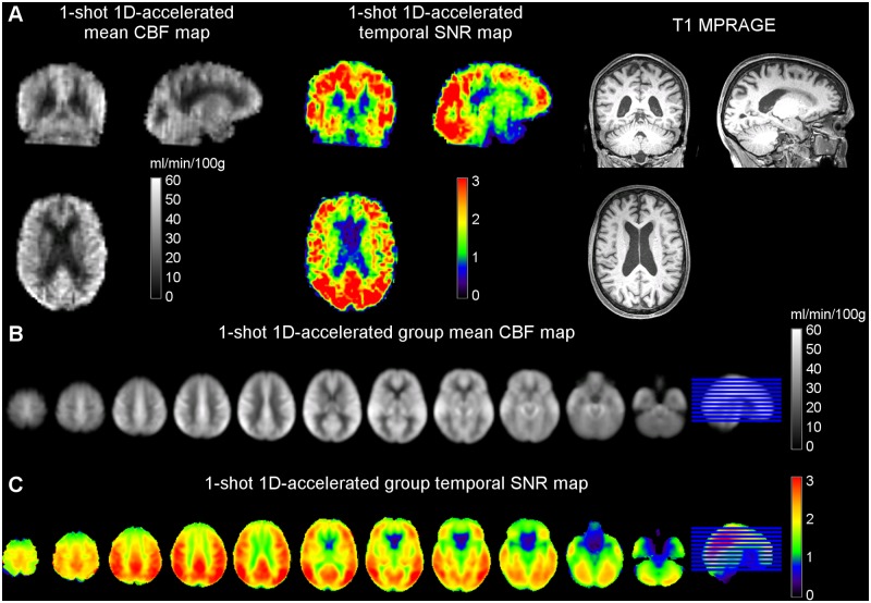
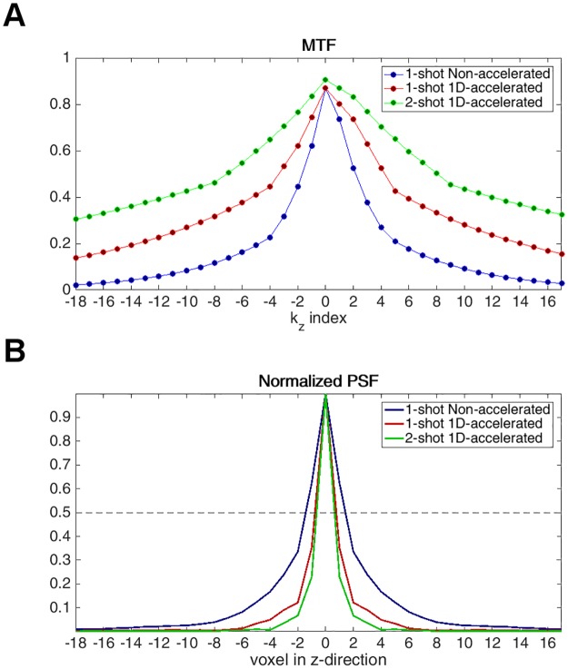
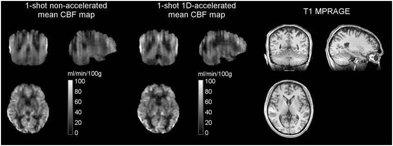
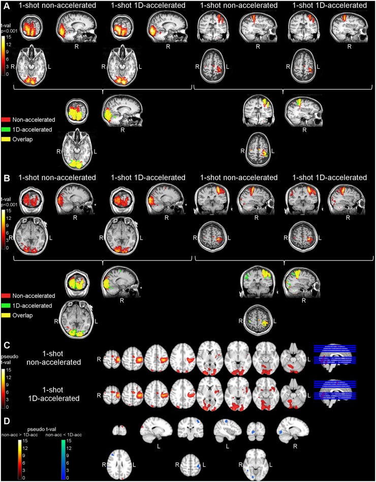
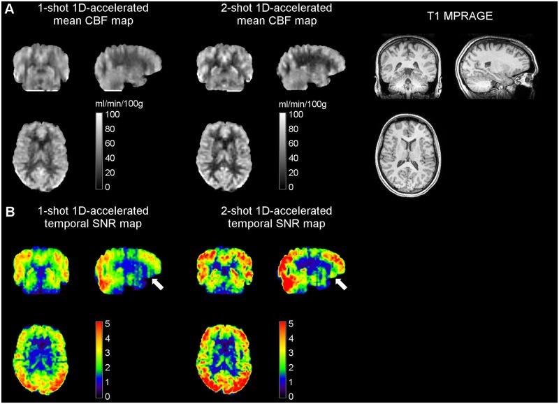
Arterial Spin Labeled (ASL) perfusion MRI enables non-invasive, quantitative measurements of tissue perfusion, and has a broad range of applications including brain functional imaging. However, ASL suffers from low signal-to-noise ratio (SNR), limiting image resolution. Acquisitions using 3D readouts are optimal for background-suppression of static signals, but can be SAR intensive and typically suffer from through-plane blurring. In this study, we investigated the use of accelerated 3D readouts to obtain whole-brain, high-SNR ASL perfusion maps and reduce SAR deposition. Parallel imaging was implemented along the partition-encoding direction in a pseudo-continuous ASL sequence with background-suppression and 3D RARE Stack-Of-Spirals readout, and its performance was evaluated in three small cohorts. First, both non-accelerated and two-fold accelerated single-shot versions of the sequence were evaluated in healthy volunteers during a motor-photic task, and the performance was compared in terms of temporal SNR, GM-WM contrast, and statistical significance of the detected activation. Secondly, single-shot 1D-accelerated imaging was compared to a two-shot accelerated version to assess benefits of SNR and spatial resolution for applications in which temporal resolution is not paramount. Third, the efficacy of this approach in clinical populations was assessed by applying the single-shot 1D-accelerated version to a larger cohort of elderly volunteers. Accelerated data demonstrated the ability to detect functional activation at the subject level, including cerebellar activity, without loss in the perfusion signal temporal stability and the statistical power of the activations. The use of acceleration also resulted in increased GM-WM contrast, likely due to reduced through-plane partial volume effects, that were further attenuated with the use of two-shot readouts. In a clinical cohort, image quality remained excellent, and expected effects of age and sex on cerebral blood flow could be detected. The sequence is freely available upon request for academic use and could benefit a broad range of cognitive and clinical neuroscience research.
动脉自旋标记(ASL)灌注磁共振成像能够对组织灌注进行无创、定量测量,并且具有广泛的应用,包括脑功能成像。然而,ASL存在信噪比(SNR)低的问题,限制了图像分辨率。使用3D读出的采集对于静态信号的背景抑制是最优的,但可能会有较高的比吸收率(SAR),并且通常会受到层面内模糊的影响。在本研究中,我们研究了使用加速3D读出以获得全脑、高SNR的ASL灌注图并减少SAR沉积。在具有背景抑制和3D RARE螺旋堆叠读出的伪连续ASL序列中,沿着分区编码方向实施并行成像,并在三个小队列中评估其性能。首先,在健康志愿者执行运动-光刺激任务期间,对该序列的非加速和两倍加速单激发版本进行了评估,并在时间SNR、灰质-白质对比度以及检测到的激活的统计学显著性方面比较了性能。其次,将单激发1D加速成像与双激发加速版本进行比较,以评估在时间分辨率不是至关重要的应用中SNR和空间分辨率的优势。第三,通过将单激发1D加速版本应用于更大的老年志愿者队列,评估了该方法在临床人群中的有效性。加速后的数据显示能够在个体水平检测功能激活,包括小脑活动,而不会损失灌注信号的时间稳定性和激活的统计功效。加速的使用还导致灰质-白质对比度增加,这可能是由于层面内部分容积效应的减少,而使用双激发读出进一步减弱了这种效应。在临床队列中,图像质量仍然优异,并且可以检测到年龄和性别对脑血流量的预期影响。该序列可应学术使用要求免费获取,可能会使广泛的认知和临床神经科学研究受益。