• 文献检索
  • 文档翻译
  • 深度研究
  • 学术资讯
  • Suppr Zotero 插件Zotero 插件
  • 邀请有礼
  • 套餐&价格
  • 历史记录
应用&插件
Suppr Zotero 插件Zotero 插件浏览器插件Mac 客户端Windows 客户端微信小程序
定价
高级版会员购买积分包购买API积分包
服务
文献检索文档翻译深度研究API 文档MCP 服务
关于我们
关于 Suppr公司介绍联系我们用户协议隐私条款
关注我们

Suppr 超能文献

核心技术专利:CN118964589B侵权必究
粤ICP备2023148730 号-1Suppr @ 2026

文献检索

告别复杂PubMed语法,用中文像聊天一样搜索,搜遍4000万医学文献。AI智能推荐,让科研检索更轻松。

立即免费搜索

文件翻译

保留排版,准确专业,支持PDF/Word/PPT等文件格式,支持 12+语言互译。

免费翻译文档

深度研究

AI帮你快速写综述,25分钟生成高质量综述,智能提取关键信息,辅助科研写作。

立即免费体验

急性脑积水的发展不会改变成年大鼠的脑组织力学特性,但会改变幼年大鼠的脑组织力学特性。

Development of acute hydrocephalus does not change brain tissue mechanical properties in adult rats, but in juvenile rats.

作者信息

Pong Alice C, Jugé Lauriane, Bilston Lynne E, Cheng Shaokoon

机构信息

Neuroscience Research Australia, Margarete Ainsworth Building, Barker Street, Randwick, Sydney, NSW, Australia.

University of New South Wales, School of Medical Sciences, Wallace Wurth Building, Kensington, Sydney, NSW Australia.

出版信息

PLoS One. 2017 Aug 24;12(8):e0182808. doi: 10.1371/journal.pone.0182808. eCollection 2017.

DOI:10.1371/journal.pone.0182808
PMID:28837671
原文链接:https://pmc.ncbi.nlm.nih.gov/articles/PMC5570303/
Abstract

INTRODUCTION

Regional changes in brain stiffness were previously demonstrated in an experimental obstructive hydrocephalus juvenile rat model. The open cranial sutures in the juvenile rats have influenced brain compression and mechanical properties during hydrocephalus development and the extent by which closed cranial sutures in adult hydrocephalic rat models affect brain stiffness in-vivo remains unclear. The aims of this study were to determine changes in brain tissue mechanical properties and brain structure size during hydrocephalus development in adult rat with fixed cranial volume and how these changes were related to brain tissue deformation.

METHODS

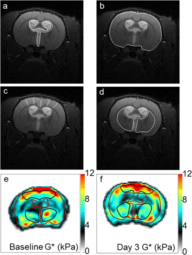
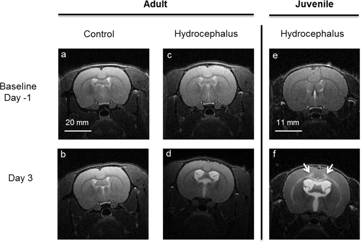
Hydrocephalus was induced in 9 female ten weeks old Sprague-Dawley rats by injecting 60 μL of a kaolin suspension (25%) into the cisterna magna under anaesthesia. 6 sham-injected age-matched female SD rats were used as controls. MR imaging (9.4T, Bruker) was performed 1 day before and then at 3 days post injection. T2-weighted anatomical MR images were collected to quantify ventricle and brain tissue cross-sectional areas. MR elastography (800 Hz) was used to measure the brain stiffness (G*, shear modulus).

RESULTS

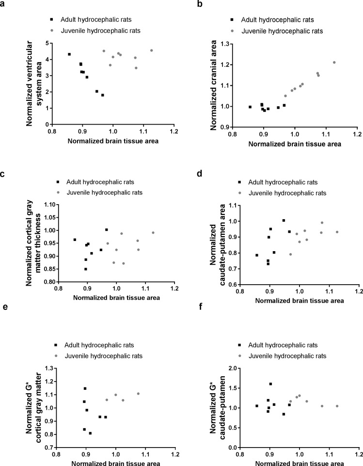
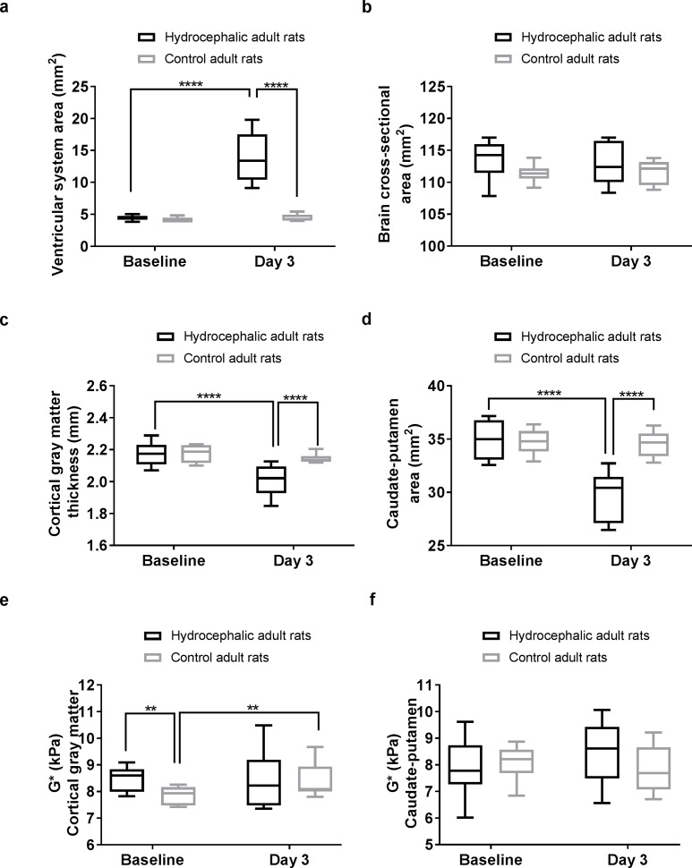
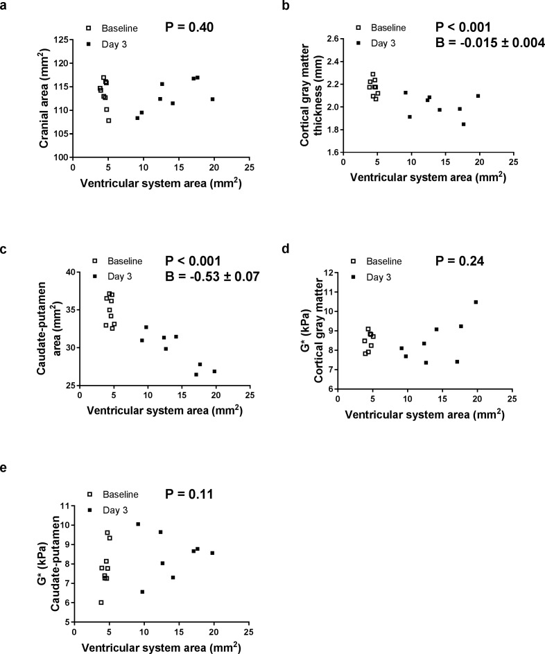
Brain tissue in the adult hydrocephalic rats was more compressed than the juvenile hydrocephalic rats because the skulls of the adult hydrocephalic rats were unable to expand like the juvenile rats. In the adult hydrocephalic rats, the cortical gray matter thickness and the caudate-putamen cross-sectional area decreased (Spearman, P < 0.001 for both) but there were no significant changes in cranial cross-sectional area (Spearman, P = 0.35), cortical gray matter stiffness (Spearman, P = 0.24) and caudate-putamen (Spearman, P = 0.11) stiffness. No significant changes in the size of brain structures were observed in the controls.

CONCLUSIONS

This study showed that although brain tissue in the adult hydrocephalic rats was severely compressed, their brain tissue stiffness did not change significantly. These results are in contrast with our previous findings in juvenile hydrocephalic rats which had significantly less brain compression (as the brain circumference was able to stretch with the cranium due to the open skull sutures) and had a significant increase in caudate putamen stiffness. These results suggest that change in brain mechanical properties in hydrocephalus is complex and is not solely dependent on brain tissue deformation. Further studies on the interactions between brain tissue stiffness, deformation, tissue oedema and neural damage are necessary before MRE can be used as a tool to track changes in brain biomechanics in hydrocephalus.

摘要

引言

先前在实验性梗阻性脑积水幼年大鼠模型中已证实脑硬度存在区域变化。幼年大鼠开放的颅骨缝在脑积水发展过程中影响了脑压缩和力学性能,而成年脑积水大鼠模型中闭合的颅骨缝在体内对脑硬度的影响程度仍不清楚。本研究的目的是确定固定颅腔容积的成年大鼠在脑积水发展过程中脑组织力学性能和脑结构大小的变化,以及这些变化与脑组织变形之间的关系。

方法

对9只10周龄雌性斯普拉格-道利大鼠在麻醉下经枕大池注射60μL高岭土悬浮液(25%)诱导脑积水。6只年龄匹配的假注射雌性SD大鼠作为对照。在注射前1天和注射后3天进行磁共振成像(9.4T,布鲁克)。采集T2加权解剖磁共振图像以量化脑室和脑组织横截面积。磁共振弹性成像(800Hz)用于测量脑硬度(G*,剪切模量)。

结果

成年脑积水大鼠的脑组织比幼年脑积水大鼠受到更严重的压缩,因为成年脑积水大鼠的颅骨不能像幼年大鼠那样扩张。在成年脑积水大鼠中,皮质灰质厚度和尾状核-壳核横截面积减小(两者的斯皮尔曼相关系数,P均<0.001),但颅横截面积(斯皮尔曼相关系数,P = 0.35)、皮质灰质硬度(斯皮尔曼相关系数,P = 0.24)和尾状核-壳核硬度(斯皮尔曼相关系数,P = 0.11)无显著变化。对照组脑结构大小无显著变化。

结论

本研究表明,尽管成年脑积水大鼠的脑组织受到严重压缩,但其脑组织硬度没有显著变化。这些结果与我们先前在幼年脑积水大鼠中的发现相反,幼年脑积水大鼠的脑压缩明显较小(由于开放的颅骨缝,脑周长能够随颅骨伸展),且尾状核-壳核硬度显著增加。这些结果表明,脑积水时脑力学性能的变化很复杂,并不完全取决于脑组织变形。在磁共振弹性成像能够用作追踪脑积水时脑生物力学变化的工具之前,有必要进一步研究脑组织硬度、变形、组织水肿和神经损伤之间的相互作用。

https://cdn.ncbi.nlm.nih.gov/pmc/blobs/cf6b/5570303/31fb3eaee413/pone.0182808.g005.jpg
https://cdn.ncbi.nlm.nih.gov/pmc/blobs/cf6b/5570303/1773af7e9884/pone.0182808.g001.jpg
https://cdn.ncbi.nlm.nih.gov/pmc/blobs/cf6b/5570303/be56c01f1b32/pone.0182808.g002.jpg
https://cdn.ncbi.nlm.nih.gov/pmc/blobs/cf6b/5570303/aa5b78605868/pone.0182808.g003.jpg
https://cdn.ncbi.nlm.nih.gov/pmc/blobs/cf6b/5570303/220b86e126af/pone.0182808.g004.jpg
https://cdn.ncbi.nlm.nih.gov/pmc/blobs/cf6b/5570303/31fb3eaee413/pone.0182808.g005.jpg

相似文献

1
Development of acute hydrocephalus does not change brain tissue mechanical properties in adult rats, but in juvenile rats.急性脑积水的发展不会改变成年大鼠的脑组织力学特性,但会改变幼年大鼠的脑组织力学特性。
PLoS One. 2017 Aug 24;12(8):e0182808. doi: 10.1371/journal.pone.0182808. eCollection 2017.
2
Changes in Rat Brain Tissue Microstructure and Stiffness during the Development of Experimental Obstructive Hydrocephalus.实验性梗阻性脑积水发展过程中大鼠脑组织微观结构和硬度的变化
PLoS One. 2016 Feb 5;11(2):e0148652. doi: 10.1371/journal.pone.0148652. eCollection 2016.
3
Characterization of juvenile and young adult mice following induction of hydrocephalus with kaolin.用高岭土诱导脑积水后幼年和青年小鼠的特征描述。
Exp Neurol. 2009 Sep;219(1):187-96. doi: 10.1016/j.expneurol.2009.05.015. Epub 2009 May 19.
4
Gray matter metabolism in acute and chronic hydrocephalus.急性和慢性脑积水时的灰质代谢
Neuroscience. 2009 Mar 17;159(2):570-7. doi: 10.1016/j.neuroscience.2009.01.008. Epub 2009 Jan 10.
5
Altered diffusion and perfusion in hydrocephalic rat brain: a magnetic resonance imaging analysis.脑积水大鼠脑内扩散与灌注的改变:磁共振成像分析
J Neurosurg. 2000 Mar;92(3):442-7. doi: 10.3171/jns.2000.92.3.0442.
6
Intracranial biomechanics of acute experimental hydrocephalus in live rats.活体大鼠急性实验性脑积水的颅内生物力学。
Neurosurgery. 2012 Nov;71(5):1032-40. doi: 10.1227/NEU.0b013e3182690a0c.
7
Magnetic resonance imaging study of extracellular fluid tracer movement in brains of immature rats with hydrocephalus.脑积水幼鼠脑内细胞外液示踪剂移动的磁共振成像研究
Neurol Res. 2000 Jan;22(1):111-6. doi: 10.1080/01616412.2000.11741045.
8
Reduced subventricular zone proliferation and white matter damage in juvenile ferrets with kaolin-induced hydrocephalus.高岭土诱导脑积水幼雪貂脑室下区增殖减少和白质损伤。
Exp Neurol. 2013 Oct;248:112-28. doi: 10.1016/j.expneurol.2013.06.004. Epub 2013 Jun 12.
9
Cerebral metabolism in experimental hydrocephalus: an in vivo 1H and 31P magnetic resonance spectroscopy study.实验性脑积水的脑代谢:一项体内氢质子和磷-31磁共振波谱研究
J Neurosurg. 1999 Oct;91(4):660-8. doi: 10.3171/jns.1999.91.4.0660.
10
The dynamics of brain and cerebrospinal fluid growth in normal versus hydrocephalic mice.正常小鼠与脑积水小鼠大脑和脑脊液生长的动态变化
J Neurosurg Pediatr. 2010 Jul;6(1):1-10. doi: 10.3171/2010.4.PEDS1014.

引用本文的文献

1
Role of Brain Elastography in the Neonatal Setting: State of the Art of Ultrasonographic Techniques and Future Perspectives.脑弹性成像在新生儿领域的作用:超声技术的现状与未来展望
Children (Basel). 2024 Jun 21;11(7):752. doi: 10.3390/children11070752.
2
Acute brain slice elastic modulus decreases over time.脑片弹性模量随时间逐渐降低。
Sci Rep. 2023 Aug 7;13(1):12826. doi: 10.1038/s41598-023-40074-z.
3
The combined use of DTI and MR elastography for monitoring microstructural changes in the developing brain of a neurodevelopmental disorder model: Poly (I:C)-induced maternal immune-activated rats.

本文引用的文献

1
Abnormal Injury Response in Spontaneous Mild Ventriculomegaly Wistar Rat Brains: A Pathological Correlation Study of Diffusion Tensor and Magnetization Transfer Imaging in Mild Traumatic Brain Injury.自发性轻度脑室扩大的Wistar大鼠脑内异常损伤反应:轻度创伤性脑损伤中扩散张量成像与磁化传递成像的病理相关性研究
J Neurotrauma. 2017 Jan 1;34(1):248-256. doi: 10.1089/neu.2015.4355. Epub 2016 Apr 11.
2
Changes in Rat Brain Tissue Microstructure and Stiffness during the Development of Experimental Obstructive Hydrocephalus.实验性梗阻性脑积水发展过程中大鼠脑组织微观结构和硬度的变化
PLoS One. 2016 Feb 5;11(2):e0148652. doi: 10.1371/journal.pone.0148652. eCollection 2016.
3
DTI 和 MR 弹性成像联合用于监测神经发育障碍模型发育中大脑的微观结构变化:聚(I:C)诱导的母体免疫激活大鼠。
PLoS One. 2023 Jan 13;18(1):e0280498. doi: 10.1371/journal.pone.0280498. eCollection 2023.
4
Brain contrast-enhanced ultrasonography and elastography in infants.婴儿脑部对比增强超声检查与弹性成像
Ultrasonography. 2022 Oct;41(4):633-649. doi: 10.14366/usg.21224. Epub 2022 Mar 30.
5
Pain Modulation from the Locus Coeruleus in a Model of Hydrocephalus: Searching for Oxidative Stress-Induced Noradrenergic Neuroprotection.脑积水中蓝斑核的疼痛调制:寻找氧化应激诱导的去甲肾上腺素能神经保护。
Int J Mol Sci. 2022 Apr 2;23(7):3970. doi: 10.3390/ijms23073970.
6
Mechanical regulation of oligodendrocyte biology.少突胶质细胞生物学的机械调控
Neurosci Lett. 2020 Jan 19;717:134673. doi: 10.1016/j.neulet.2019.134673. Epub 2019 Dec 12.
7
VEGF Signaling in Neurological Disorders.血管内皮生长因子信号通路在神经疾病中的作用
Int J Mol Sci. 2018 Jan 17;19(1):275. doi: 10.3390/ijms19010275.
Microvasculature alters the dispersion properties of shear waves--a multi-frequency MR elastography study.
微血管改变剪切波的弥散特性——一项多频磁共振弹性成像研究。
NMR Biomed. 2015 Dec;28(12):1763-71. doi: 10.1002/nbm.3438.
4
MR Elastography Demonstrates Increased Brain Stiffness in Normal Pressure Hydrocephalus.磁共振弹性成像显示正常压力脑积水患者脑硬度增加。
AJNR Am J Neuroradiol. 2016 Mar;37(3):462-7. doi: 10.3174/ajnr.A4560. Epub 2015 Nov 5.
5
MR Elastography Can Be Used to Measure Brain Stiffness Changes as a Result of Altered Cranial Venous Drainage During Jugular Compression.磁共振弹性成像可用于测量颈静脉压迫期间因颅内静脉引流改变而导致的脑硬度变化。
AJNR Am J Neuroradiol. 2015 Oct;36(10):1971-7. doi: 10.3174/ajnr.A4361. Epub 2015 Jun 4.
6
Imaging of spontaneous ventriculomegaly and vascular malformations in Wistar rats: implications for preclinical research.Wistar大鼠自发性脑室扩大和血管畸形的影像学研究:对临床前研究的启示
J Neuropathol Exp Neurol. 2014 Dec;73(12):1152-65. doi: 10.1097/NEN.0000000000000140.
7
Combining MR elastography and diffusion tensor imaging for the assessment of anisotropic mechanical properties: a phantom study.结合磁共振弹性成像和扩散张量成像评估各向异性力学特性:一项体模研究。
J Magn Reson Imaging. 2013 Jan;37(1):217-26. doi: 10.1002/jmri.23797. Epub 2012 Sep 17.
8
Demyelination reduces brain parenchymal stiffness quantified in vivo by magnetic resonance elastography.脱髓鞘降低了磁共振弹性成像在体量化的脑实质硬度。
Proc Natl Acad Sci U S A. 2012 Apr 24;109(17):6650-5. doi: 10.1073/pnas.1200151109. Epub 2012 Apr 5.
9
Using static preload with magnetic resonance elastography to estimate large strain viscoelastic properties of bovine liver.利用磁共振弹性成像的静态预载技术估计牛肝的大应变黏弹性性质。
J Biomech. 2011 Sep 2;44(13):2461-5. doi: 10.1016/j.jbiomech.2011.06.023. Epub 2011 Jul 16.
10
Alteration of brain viscoelasticity after shunt treatment in normal pressure hydrocephalus.常压性脑积水分流治疗后脑黏弹性改变。
Neuroradiology. 2012 Mar;54(3):189-96. doi: 10.1007/s00234-011-0871-1. Epub 2011 May 3.