Kupersmith Mark J, Sibony Patrick A, Dave Sarita
New York Eye and Ear Infirmary and Icahn School of Medicine at Mount Sinai, New York, New York, United States.
Department of Ophthalmology, State University of New York at Stony Brook, Stony Brook, New York, United States.
Invest Ophthalmol Vis Sci. 2017 Aug 1;58(10):4286-4291. doi: 10.1167/iovs.17-22140.
We hypothesized that the edema/swelling in the retina due to acute nonarteritic anterior ischemic optic neuropathy (NAION) can induce retinal folds (RF). We determined the pattern and frequency of folds in NAION at presentation and in follow-up, and the relationship between folds and a number of functional and structural parameters over time.
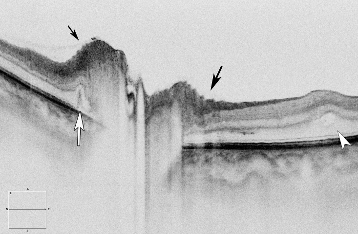
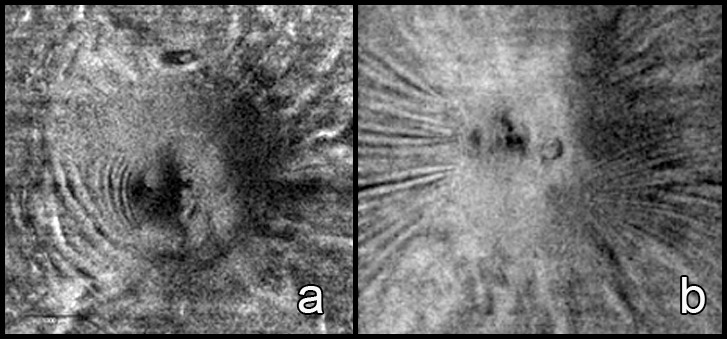
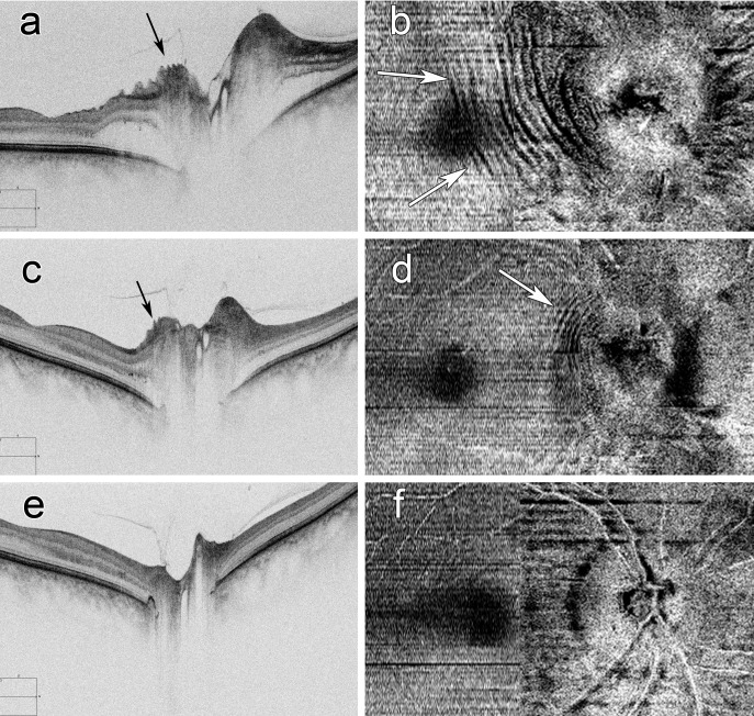
We prospectively studied eyes with acute NAION by spectral-domain optic coherence tomography (SD-OCT). We used transaxial and en face views to evaluate the presence of peripapillary fluid (PPF), peripapillary wrinkles (PPW), RF, choroidal folds (CF), creases, macular edema, and vitreous traction on the optic disc. Retinal deformations were correlated with the retinal nerve fiber layer (RNFL) thickness, logMAR visual acuity (VA) and mean deviation (MD).
At presentation, 60 eyes had mean RNFL = 224 ± 75 μm, no vitreous traction, and similar VA and MD regardless of the retinal deformation or macular edema. There was PPF in 73%, PPW in 57%, RF in 38%, creases in 20%, and macular edema in 18% of eyes, and no CF. Eyes with retinal deformations had significantly greater RNFL thickness (P< 0.026). At 1 to 2 months, 49 eyes had reduction of the RNFL (112 ± 40 μm, P = 0.001) and unchanged VA and MD that did not correlate with fewer eyes having PPF (15%, P = 0.001), PPW (10%, P = 0.001), RF (10%, P = 0.001), creases (17%), and macular edema (0%, P = 0.007).
RF in NAION reflect stresses and strains due to extracellular fluid without increased pressure in the retrolaminar tissue and subarachnoid space, seen with papilledema. In NAION, the deformations and their resolution do not correlate with vision loss.
我们推测急性非动脉炎性前部缺血性视神经病变(NAION)所致视网膜水肿/肿胀可诱发视网膜皱褶(RF)。我们确定了NAION患者初诊及随访时皱褶的形态和频率,以及随时间推移皱褶与一些功能和结构参数之间的关系。
我们采用光谱域光学相干断层扫描(SD - OCT)对急性NAION患者的眼睛进行前瞻性研究。我们使用横断面和正面视图评估视乳头周围液体(PPF)、视乳头周围皱纹(PPW)、RF、脉络膜皱褶(CF)、皱痕、黄斑水肿以及视盘上的玻璃体牵拉情况。视网膜变形与视网膜神经纤维层(RNFL)厚度、对数最小分辨角视力(logMAR VA)及平均偏差(MD)相关。
初诊时,60只眼的平均RNFL为224±75μm,无玻璃体牵拉,无论视网膜变形或黄斑水肿情况如何,视力和MD相似。73%的眼存在PPF,57%存在PPW,38%存在RF,20%存在皱痕,18%存在黄斑水肿,无CF。存在视网膜变形的眼RNFL厚度显著更大(P < 0.026)。在1至2个月时,49只眼的RNFL变薄(112±40μm,P = 0.001),视力和MD未变,且PPF(15%,P = 0.001)、PPW(10%,P = 0.001)、RF(10%,P = 0.001)、皱痕(17%)及黄斑水肿(0%,P = 0.007)出现的眼数减少与之无关。
NAION中的RF反映了细胞外液引起的应力和应变,而非如视乳头水肿时在视网膜后组织和蛛网膜下腔压力升高所致。在NAION中,变形及其消退与视力丧失无关。