Department of Medicine, Vanderbilt University School of Medicine, Nashville, Tennessee, USA; Medical and Research Services, Department of Veterans Affairs Hospital, Tennessee Valley Healthcare System, Nashville, Tennessee, USA.
Section of Pediatric Nephrology, Department of Pediatrics, Tulane University Health Sciences Center, New Orleans, Louisiana, USA; The Hypertension and Renal Centers of Excellence, Tulane University Health Sciences Center, New Orleans, Louisiana, USA.
Kidney Int. 2017 Dec;92(6):1370-1383. doi: 10.1016/j.kint.2017.06.015. Epub 2017 Aug 26.
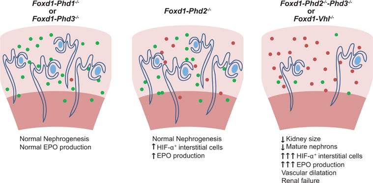
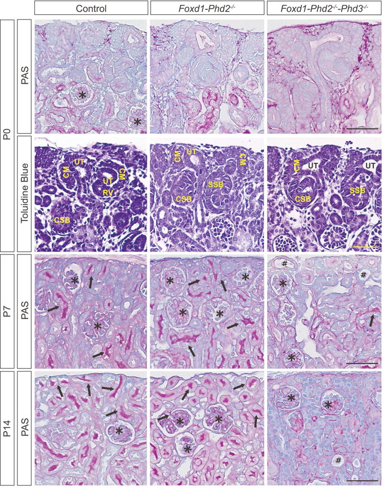
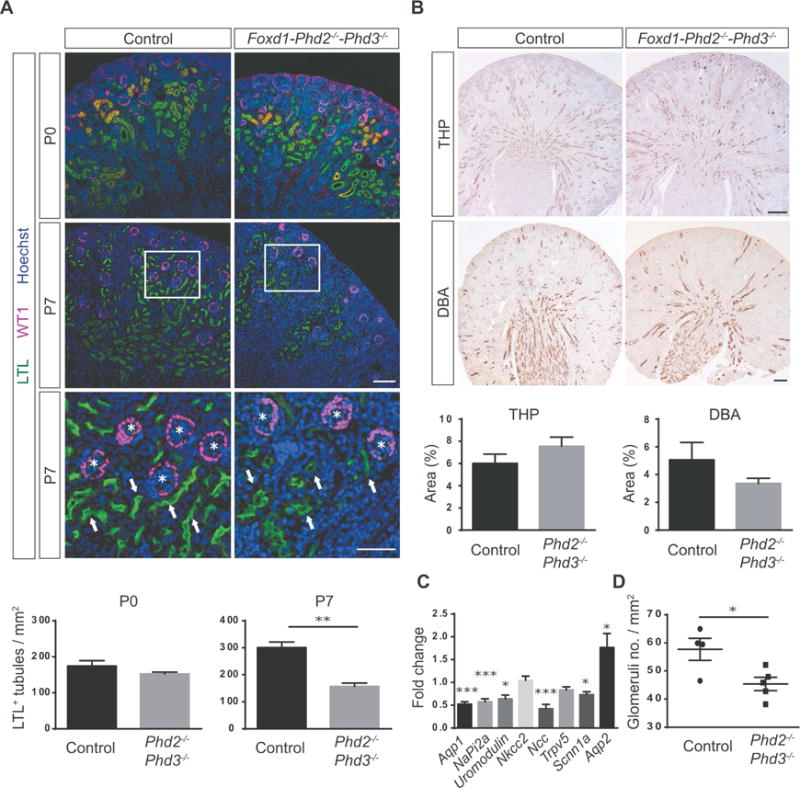
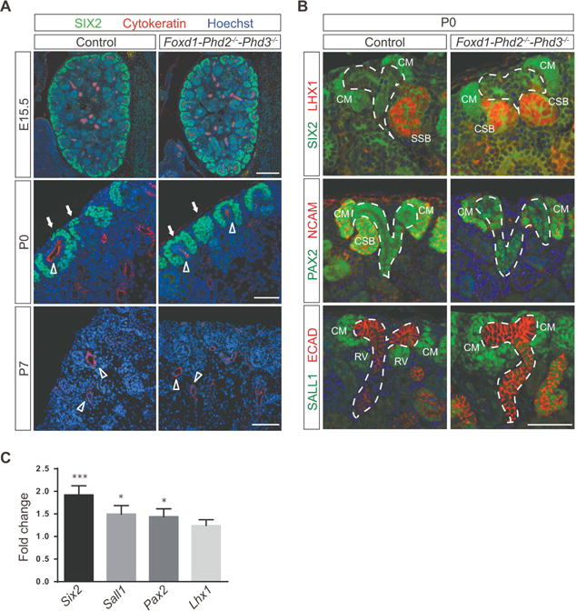
Hypoxia in the embryo is a frequent cause of intra-uterine growth retardation, low birth weight, and multiple organ defects. In the kidney, this can lead to low nephron endowment, predisposing to chronic kidney disease and arterial hypertension. A key component in cellular adaptation to hypoxia is the hypoxia-inducible factor pathway, which is regulated by prolyl-4-hydroxylase domain (PHD) dioxygenases PHD1, PHD2, and PHD3. In the adult kidney, PHD oxygen sensors are differentially expressed in a cell type-dependent manner and control the production of erythropoietin in interstitial cells. However, the role of interstitial cell PHDs in renal development has not been examined. Here we used a genetic approach in mice to interrogate PHD function in FOXD1-expressing stroma during nephrogenesis. We demonstrate that PHD2 and PHD3 are essential for normal kidney development as the combined inactivation of stromal PHD2 and PHD3 resulted in renal failure that was associated with reduced kidney size, decreased numbers of glomeruli, and abnormal postnatal nephron formation. In contrast, nephrogenesis was normal in animals with individual PHD inactivation. We furthermore demonstrate that the defect in nephron formation in PHD2/PHD3 double mutants required intact hypoxia-inducible factor-2 signaling and was dependent on the extent of stromal hypoxia-inducible factor activation. Thus, hypoxia-inducible factor prolyl-4-hydroxylation in renal interstitial cells is critical for normal nephron formation.
胚胎缺氧是宫内生长迟缓、低出生体重和多种器官缺陷的常见原因。在肾脏中,这可能导致肾单位数量不足,易患慢性肾脏病和动脉高血压。细胞适应缺氧的关键组成部分是缺氧诱导因子通路,该通路受脯氨酰-4-羟化酶结构域(PHD)双加氧酶 PHD1、PHD2 和 PHD3 的调节。在成年肾脏中,PHD 氧传感器以细胞类型依赖的方式差异表达,并控制间质细胞中促红细胞生成素的产生。然而,间质细胞 PHD 在肾脏发育中的作用尚未被研究。在这里,我们使用小鼠中的遗传方法来研究在肾发生过程中 FOXD1 表达的基质中 PHD 的功能。我们证明 PHD2 和 PHD3 对于正常肾脏发育是必不可少的,因为基质 PHD2 和 PHD3 的联合失活导致肾功能衰竭,伴有肾脏缩小、肾小球数量减少和出生后肾小球形成异常。相比之下,单个 PHD 失活的动物的肾发生正常。我们还证明,在 PHD2/PHD3 双突变体中,肾单位形成的缺陷需要完整的缺氧诱导因子-2 信号通路,并且依赖于基质缺氧诱导因子激活的程度。因此,肾脏间质细胞中缺氧诱导因子脯氨酰-4-羟化对正常肾单位形成至关重要。