Bristol Renal, Bristol Medical School, University of Bristol, Whitson Street, Bristol, BS1 3NY, UK.
Global Research, Novo Nordisk A/S, Måløv, Denmark.
Diabetologia. 2017 Nov;60(11):2299-2311. doi: 10.1007/s00125-017-4394-0. Epub 2017 Aug 29.
AIMS/HYPOTHESIS: Podocytes are insulin-responsive cells of the glomerular filtration barrier and are key in preventing albuminuria, a hallmark feature of diabetic nephropathy. While there is evidence that a loss of insulin signalling to podocytes is detrimental, the molecular mechanisms underpinning the development of podocyte insulin resistance in diabetes remain unclear. Thus, we aimed to further investigate podocyte insulin responses early in the context of diabetic nephropathy.
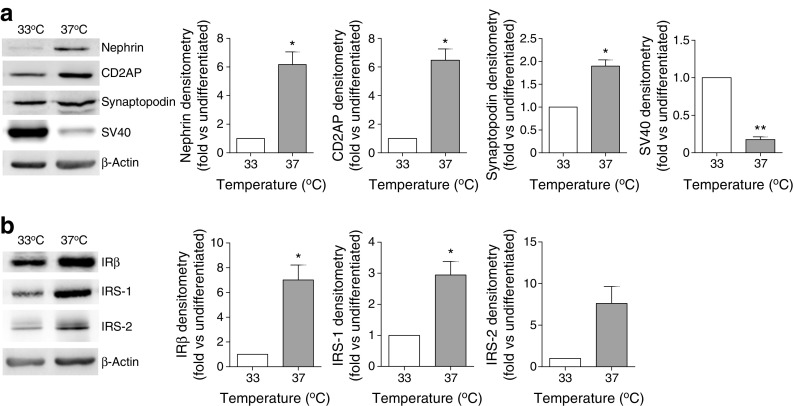
Conditionally immortalised human and mouse podocyte cell lines and glomeruli isolated from db/db DBA/2J mice were studied. Podocyte insulin responses were investigated with western blotting, cellular glucose uptake assays and automated fluorescent imaging of the actin cytoskeleton. Quantitative (q)RT-PCR was employed to investigate changes in mRNA. Human cell lines stably overproducing the insulin receptor (IR) and nephrin were also generated, using lentiviral constructs.
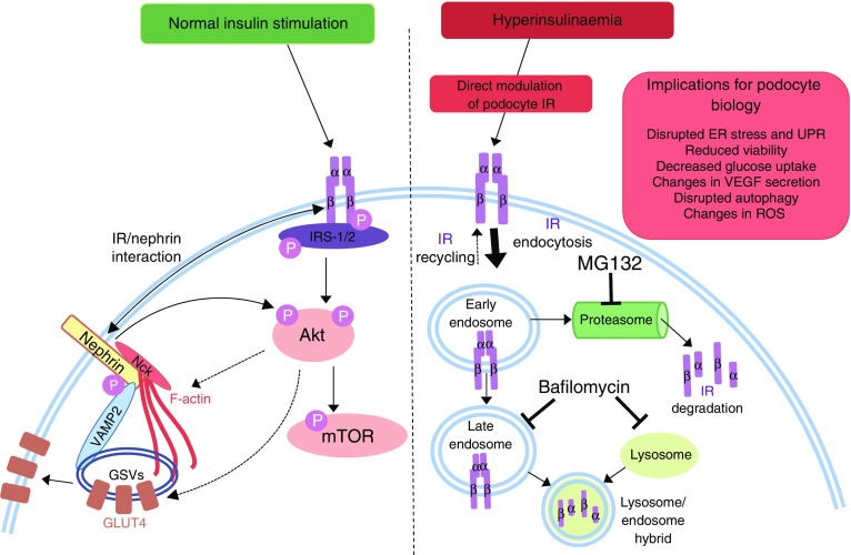
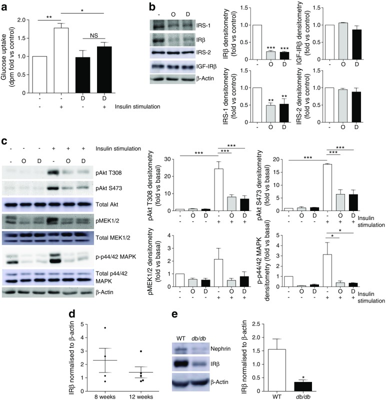
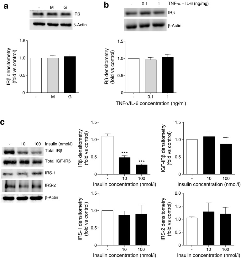
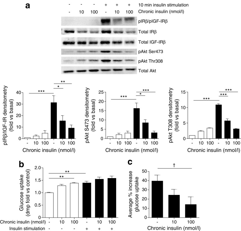
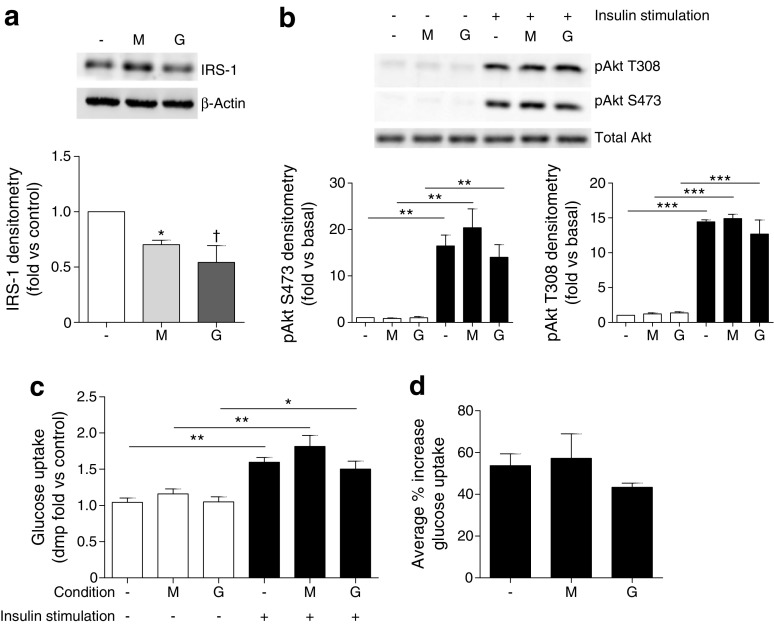
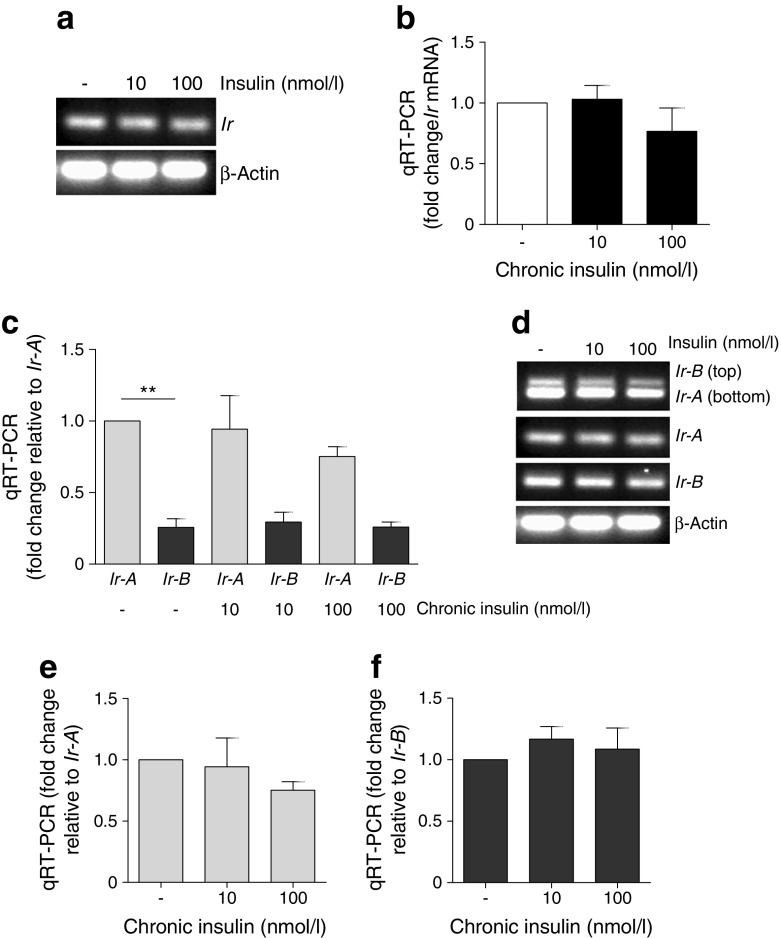
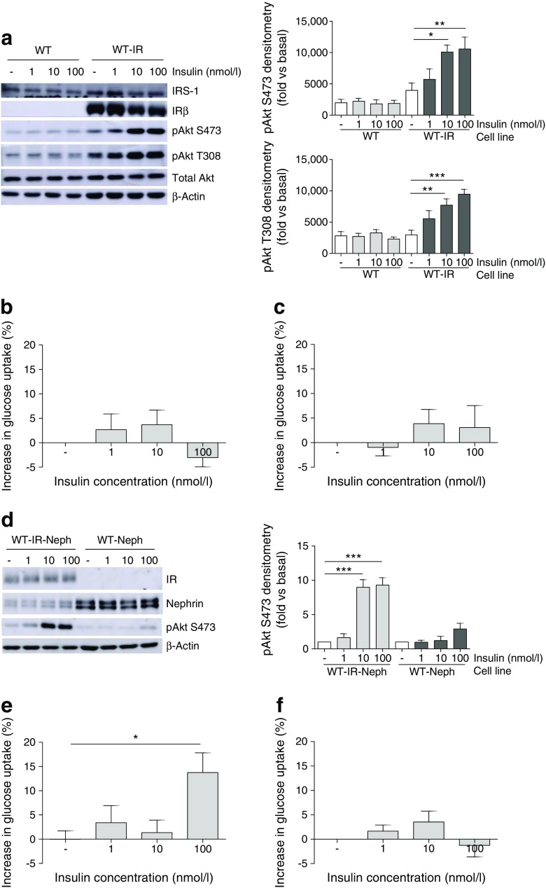
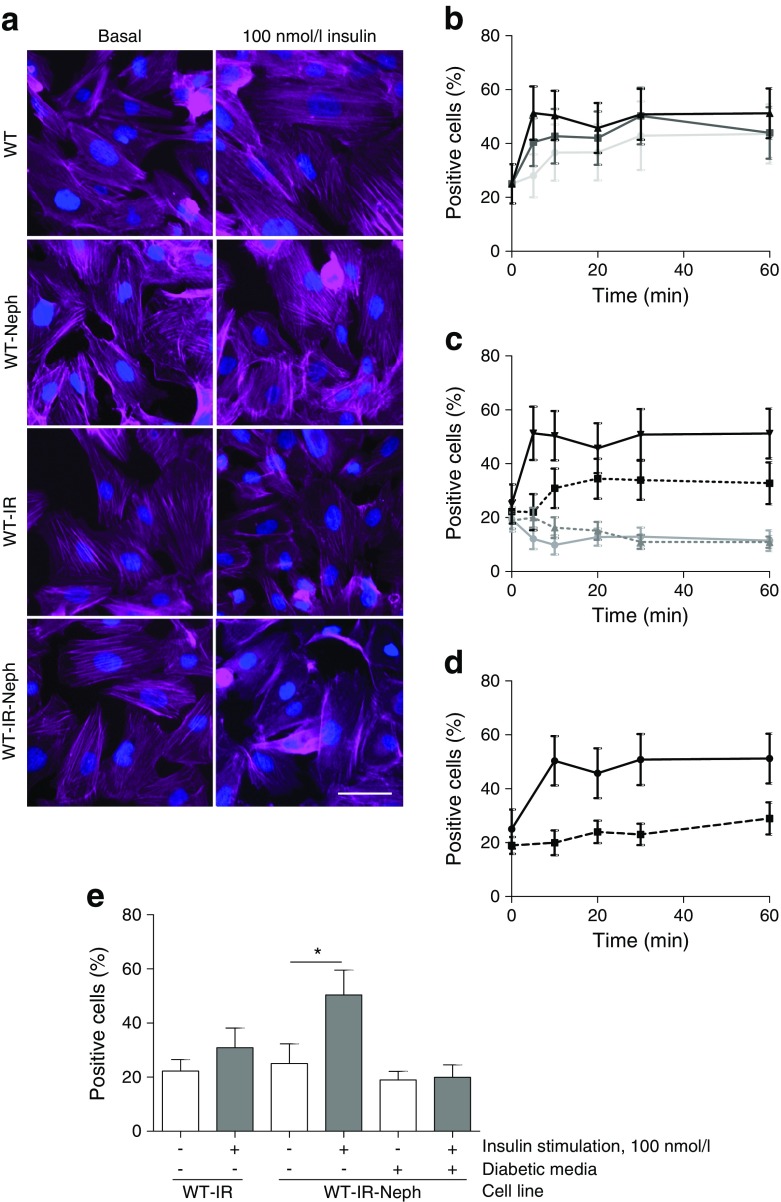
Podocytes exposed to a diabetic environment (high glucose, high insulin and the proinflammatory cytokines TNF-α and IL-6) become insulin resistant with respect to glucose uptake and activation of phosphoinositide 3-kinase (PI3K) and mitogen-activated protein kinase (MAPK) signalling. These podocytes lose expression of the IR as a direct consequence of prolonged exposure to high insulin concentrations, which causes an increase in IR protein degradation via a proteasome-dependent and bafilomycin-sensitive pathway. Reintroducing the IR into insulin-resistant human podocytes rescues upstream phosphorylation events, but not glucose uptake. Stable expression of nephrin is also required for the insulin-stimulated glucose uptake response in podocytes and for efficient insulin-stimulated remodelling of the actin cytoskeleton.
CONCLUSIONS/INTERPRETATION: Together, these results suggest that IR degradation, caused by high levels of insulin, drives early podocyte insulin resistance, and that both the IR and nephrin are required for full insulin sensitivity of this cell. This could be highly relevant for the development of nephropathy in individuals with type 2 diabetes, who are commonly hyperinsulinaemic in the early phases of their disease.
目的/假设:足细胞是肾小球滤过屏障中对胰岛素有反应的细胞,是防止白蛋白尿(糖尿病肾病的一个标志性特征)的关键。虽然有证据表明,足细胞中胰岛素信号的丧失是有害的,但糖尿病中导致足细胞胰岛素抵抗的分子机制尚不清楚。因此,我们旨在进一步研究糖尿病肾病早期阶段足细胞的胰岛素反应。
研究了条件永生化的人源和鼠源足细胞系以及来自 db/db DBA/2J 小鼠的肾小球。通过 Western blot、细胞葡萄糖摄取测定和肌动蛋白细胞骨架的自动荧光成像研究了足细胞的胰岛素反应。采用定量(q)RT-PCR 研究了 mRNA 的变化。还使用慢病毒构建体生成了稳定过表达胰岛素受体(IR)和nephrin 的人源细胞系。
暴露于高糖、高胰岛素以及促炎细胞因子 TNF-α 和 IL-6 的糖尿病环境中的足细胞对葡萄糖摄取和磷酸肌醇 3-激酶(PI3K)和丝裂原活化蛋白激酶(MAPK)信号的激活表现出胰岛素抵抗。这些足细胞由于长时间暴露于高胰岛素浓度而直接丧失 IR 的表达,这导致通过蛋白酶体依赖性和巴弗洛霉素敏感途径增加 IR 蛋白降解。将 IR 重新引入胰岛素抵抗的人足细胞中可挽救上游磷酸化事件,但不能挽救葡萄糖摄取。nephrin 的稳定表达也是足细胞中胰岛素刺激的葡萄糖摄取反应和胰岛素刺激的肌动蛋白细胞骨架重塑所必需的。
结论/解释:综上所述,这些结果表明,高胰岛素水平引起的 IR 降解导致早期足细胞胰岛素抵抗,并且 IR 和 nephrin 都是该细胞完全胰岛素敏感性所必需的。这对于 2 型糖尿病患者的肾病发展可能具有高度相关性,这些患者在疾病的早期阶段通常会出现高胰岛素血症。