AlDarrab Abdulrahman, Alrajeh Mohammed, Alsuhaibani Adel H
Department of Surgery, College of Medicine, Prince Sattam Bin Abdulaziz University, Saudi Arabia.
Department of Surgery, King Abdulaziz Medical City, Riyadh, Saudi Arabia.
Saudi J Ophthalmol. 2017 Jul-Sep;31(3):131-134. doi: 10.1016/j.sjopt.2017.05.014. Epub 2017 Jun 1.
To study the effect of posterior blepharitis on meibomian glands using infrared meibography and to correlate the results with tear film parameters.
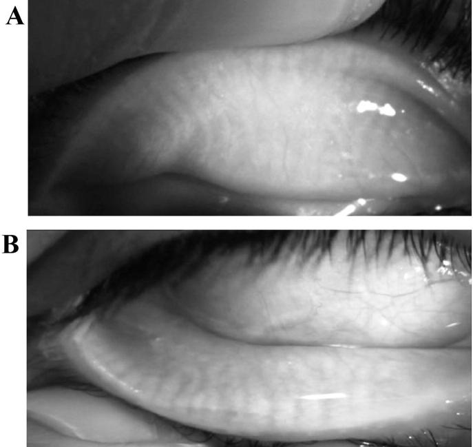
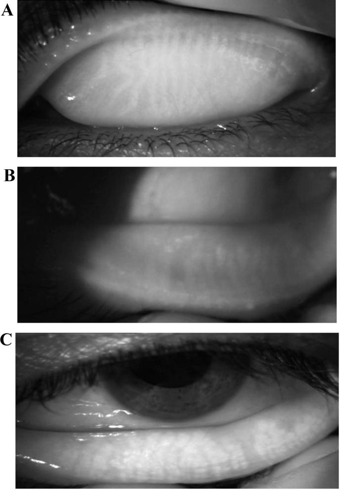
This is a prospective cohort study. The study included eyes from two groups: 86 eyes of healthy volunteers' eyes and 72 eyes with posterior blepharitis. Participants were examined, and diagnosis of posterior blepharitis was achieved clinically based on signs of posterior blepharitis. Clinical assessment of dryness was performed including slit lamp examination looking for signs of posterior blepharitis, tear breakup time (TBUT), superficial punctate keratopathy (SPK), Schirmer II test (with anesthesia) and meibum score. Non-contact meibography was performed for both upper and lower eyelids using the meibo-grade system which involved distortion of meibomian gland, shortening and dropout.
Lid margin abnormalities (Telangiectasia, lid margin swelling and hyperemia) were all significantly higher in the posterior blepharitis group. SPK, meibum score, meibography dropout, distortion, shortening, and total meibography were all significantly higher in the posterior blepharitis group as well as meibum score ( value < 0.001). TBUT was significantly shorter in the posterior blepharitis group ( value < 0.001). There was no significant difference between the two groups in Schirmer's II test.
Meibography can be a helpful non-invasive tool for the clinical evaluation of the extent of the anatomical damage in patients having meibomian glands loss due to posterior blepharitis. Knowing the extent of damage in meibomian glands may help in selecting the appropriate treatment modality and expect the response to treatment in patients with posterior blepharitis.
使用红外睑板腺造影术研究后部睑缘炎对睑板腺的影响,并将结果与泪膜参数相关联。
这是一项前瞻性队列研究。该研究纳入了两组的眼睛:86只健康志愿者的眼睛和72只患有后部睑缘炎的眼睛。对参与者进行检查,根据后部睑缘炎的体征在临床上做出后部睑缘炎的诊断。进行干眼的临床评估,包括裂隙灯检查以寻找后部睑缘炎的体征、泪膜破裂时间(TBUT)、浅层点状角膜炎(SPK)、Schirmer II试验(麻醉下)和睑脂评分。使用睑板腺分级系统对上下眼睑进行非接触式睑板腺造影,该系统涉及睑板腺变形、缩短和缺失。
后部睑缘炎组的睑缘异常(毛细血管扩张、睑缘肿胀和充血)均显著更高。后部睑缘炎组的SPK、睑脂评分、睑板腺造影缺失、变形、缩短以及总的睑板腺造影结果也均显著更高,睑脂评分(P值<0.001)。后部睑缘炎组的TBUT显著更短(P值<0.001)。两组在Schirmer II试验中无显著差异。
睑板腺造影术可以成为一种有用的非侵入性工具,用于临床评估因后部睑缘炎导致睑板腺丧失的患者的解剖学损伤程度。了解睑板腺的损伤程度可能有助于选择合适的治疗方式,并预测后部睑缘炎患者的治疗反应。