Unidad de Investigación en Neurodesarrollo, Departamento de Neurobiología Conductual y Cognitiva, Instituto de Neurobiología, Universidad Nacional Autónoma de México (UNAM), Campus Juriquilla, Mexico.
USC Mark and Mary Stevens Neuroimaging and Informatics Institute, 2025 Zonal Avenue, SHN, Los Angeles, California 90033, USA.
Neuroimage Clin. 2017 Aug 14;16:355-368. doi: 10.1016/j.nicl.2017.08.015. eCollection 2017.
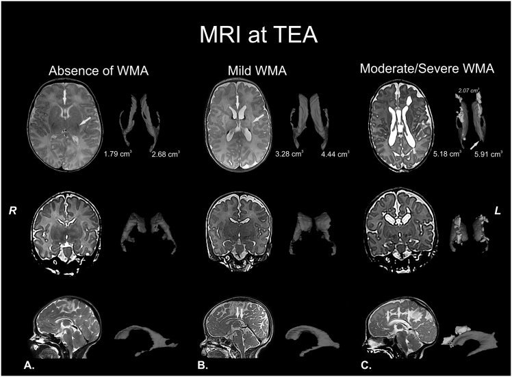
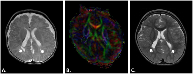
Perinatal care advances emerging over the past twenty years have helped to diminish the mortality and severe neurological morbidity of extremely and very preterm neonates (e.g., cystic Periventricular Leukomalacia [c-PVL] and Germinal Matrix Hemorrhage - Intraventricular Hemorrhage [GMH-IVH grade 3-4/4]; 22 to < 32 weeks of gestational age, GA). However, motor and/or cognitive disabilities associated with mild-to-moderate white and gray matter injury are frequently present in this population (e.g., non-cystic Periventricular Leukomalacia [non-cystic PVL], neuronal-axonal injury and GMH-IVH grade 1-2/4). Brain research studies using magnetic resonance imaging (MRI) report that 50% to 80% of extremely and very preterm neonates have diffuse white matter abnormalities (WMA) which correspond to only the minimum grade of severity. Nevertheless, mild-to-moderate diffuse WMA has also been associated with significant affectations of motor and cognitive activities. Due to increased neonatal survival and the intrinsic characteristics of diffuse WMA, there is a growing need to study the brain of the premature infant using non-invasive neuroimaging techniques sensitive to microscopic and/or diffuse lesions. This emerging need has led the scientific community to try to bridge the gap between concepts or ideas from different methodologies and approaches; for instance, neuropathology, neuroimaging and clinical findings. This is evident from the combination of intense pre-clinical and clinicopathologic research along with neonatal neurology and quantitative neuroimaging research. In the following review, we explore literature relating the most frequently observed neuropathological patterns with the recent neuroimaging findings in preterm newborns and infants with perinatal brain injury. Specifically, we focus our discussions on the use of neuroimaging to aid diagnosis, measure morphometric brain damage, and track long-term neurodevelopmental outcomes.
过去 20 年来,围产期护理的进步帮助降低了极早产儿和极早产儿(例如,囊泡性脑白质软化症[囊性 PVL]和生发基质出血-脑室内出血[GMH-IVH 3-4/4 级])的死亡率和严重神经发育障碍发生率。然而,该人群中经常存在与轻度至中度白质和灰质损伤相关的运动和/或认知障碍(例如,非囊泡性脑白质软化症[非囊性 PVL]、神经元-轴突损伤和 GMH-IVH 1-2/4 级)。使用磁共振成像(MRI)的脑研究报告称,50%至 80%的极早产儿和极早产儿有弥漫性白质异常(WMA),这些异常仅对应于最低严重程度的等级。然而,轻度至中度弥漫性 WMA 也与运动和认知活动的显著影响有关。由于新生儿存活率的提高和弥漫性 WMA 的内在特征,越来越需要使用对微观和/或弥漫性病变敏感的非侵入性神经影像学技术研究早产儿的大脑。这种新出现的需求促使科学界试图弥合来自不同方法和方法的概念或想法之间的差距;例如,神经病理学、神经影像学和临床发现。这从临床前和临床病理研究与新生儿神经病学和定量神经影像学研究的结合中可以明显看出。在以下综述中,我们探讨了与围产期脑损伤的早产儿和婴儿最近的神经影像学发现最常观察到的神经病理学模式相关的文献。具体来说,我们将讨论重点放在使用神经影像学来辅助诊断、测量形态计量脑损伤以及跟踪长期神经发育结果上。