Department of Neonatology, Hospital Sant Joan de Déu, Institut de Recerca Sant Joan de Déu, Barcelona, Spain.
Department of Neonatology, Quironsalud Madrid University Hospital and Biomedical Research Foundation, La Paz University Hospital Madrid, Madrid, Spain.
Pediatr Res. 2020 Mar;87(Suppl 1):37-49. doi: 10.1038/s41390-020-0781-1.
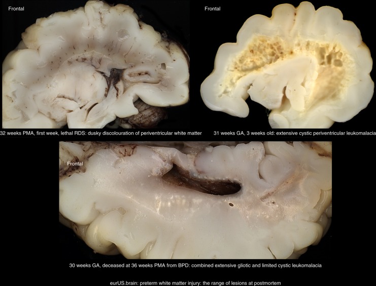
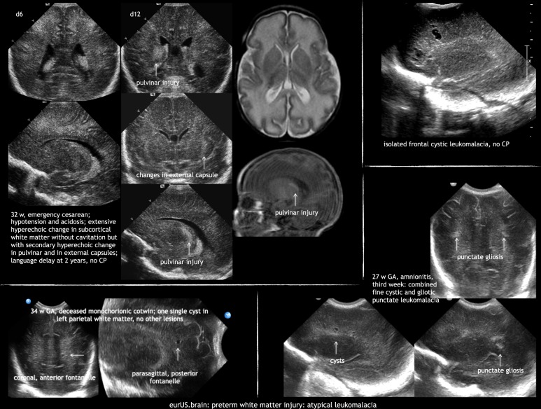
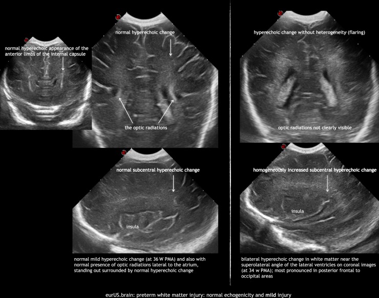
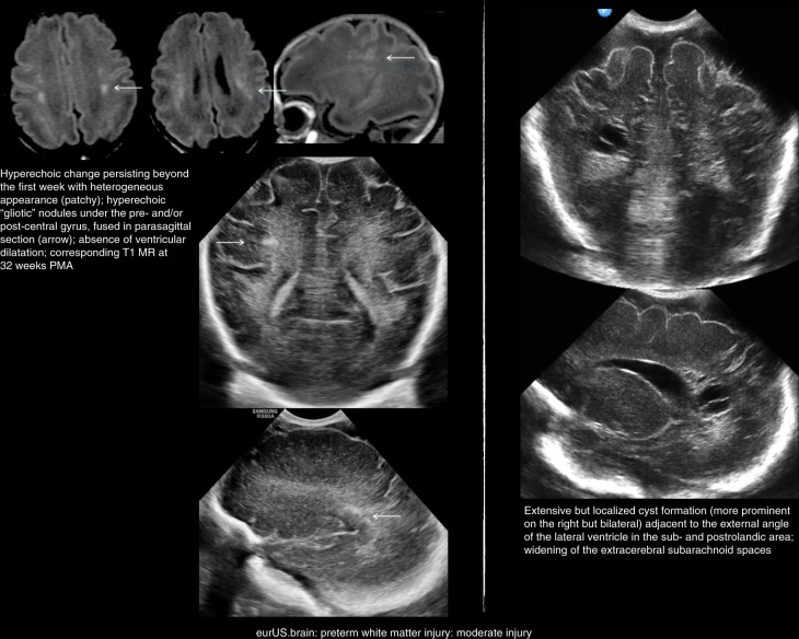
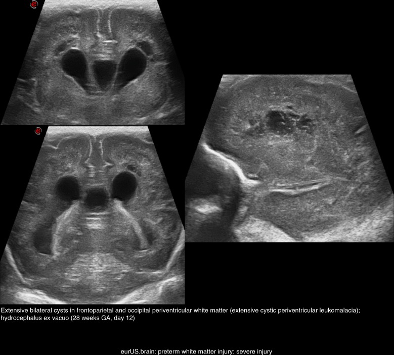
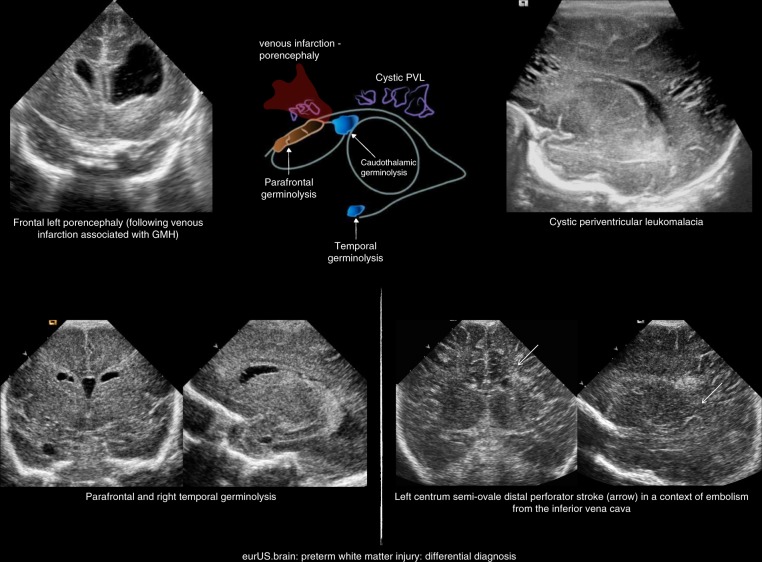
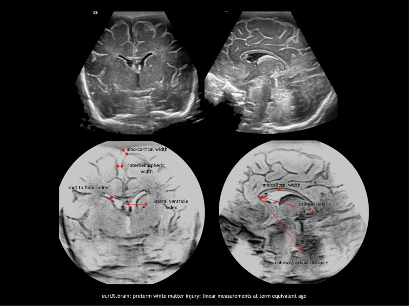
White matter injury (WMI) is the most frequent form of preterm brain injury. Cranial ultrasound (CUS) remains the preferred modality for initial and sequential neuroimaging in preterm infants, and is reliable for the diagnosis of cystic periventricular leukomalacia. Although magnetic resonance imaging is superior to CUS in detecting the diffuse and more subtle forms of WMI that prevail in very premature infants surviving nowadays, recent improvement in the quality of neonatal CUS imaging has broadened the spectrum of preterm white matter abnormalities that can be detected with this technique. We propose a structured CUS assessment of WMI of prematurity that seeks to account for both cystic and non-cystic changes, as well as signs of white matter loss and impaired brain growth and maturation, at or near term equivalent age. This novel assessment system aims to improve disease description in both routine clinical practice and clinical research. Whether this systematic assessment will improve prediction of outcome in preterm infants with WMI still needs to be evaluated in prospective studies.
脑白质损伤(WMI)是早产儿脑损伤最常见的形式。头颅超声(CUS)仍然是早产儿初始和连续神经影像学检查的首选方式,对囊性脑室周围白质软化症的诊断是可靠的。虽然磁共振成像(MRI)在检测现今存活的极早产儿中弥漫性和更细微的 WMI 方面优于 CUS,但最近新生儿 CUS 成像质量的提高拓宽了可以用该技术检测到的早产儿脑白质异常的范围。我们提出了一种针对早产儿 WMI 的结构化 CUS 评估方法,旨在同时考虑囊性和非囊性变化,以及白质损失和脑生长和成熟受损的迹象,在接近足月等效年龄时进行评估。这种新的评估系统旨在改善常规临床实践和临床研究中的疾病描述。这种系统评估是否会改善 WMI 早产儿的预后预测仍需要前瞻性研究来评估。