Wang Yushan, Sawyer Thomas W, Tse Yiu Chung, Fan Changyang, Hennes Grant, Barnes Julia, Josey Tyson, Weiss Tracy, Nelson Peggy, Wong Tak Pan
Defence Research and Development Canada, Suffield Research Centre, Medicine Hat, AB, Canada.
Department of Psychiatry, Douglas Mental Health University Institute, McGill University, Montreal, QC, Canada.
Front Neurol. 2017 Aug 18;8:413. doi: 10.3389/fneur.2017.00413. eCollection 2017.
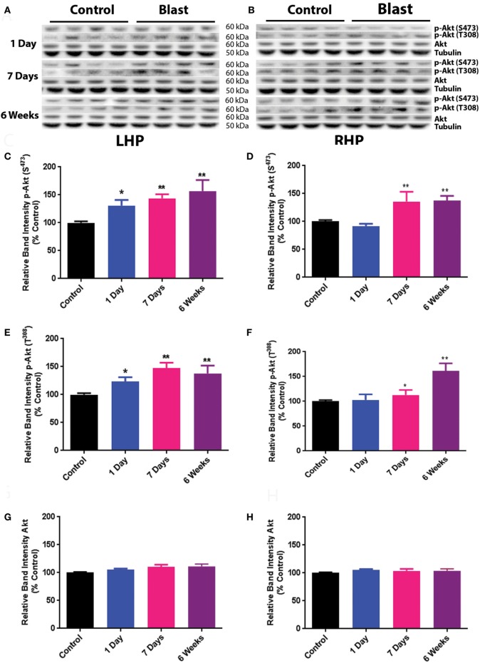
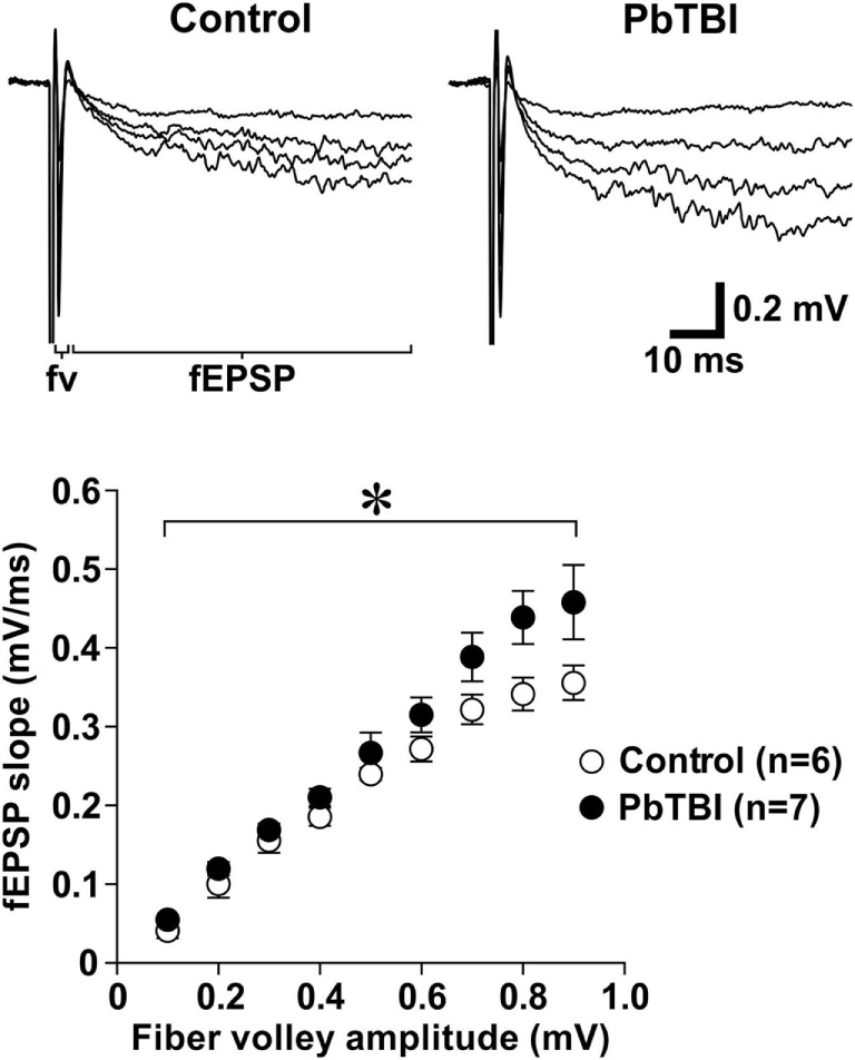
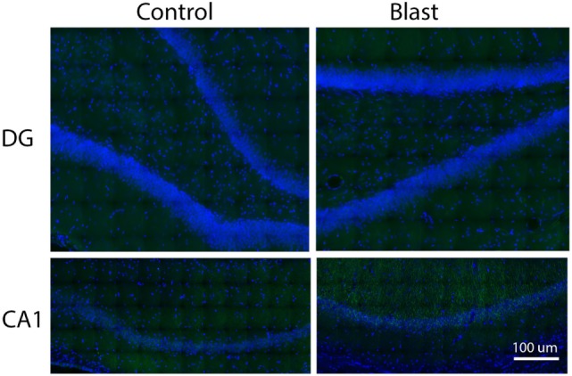
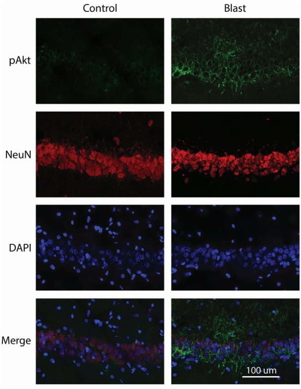
Traumatic brain injury (TBI) due to blast from improvised explosive devices has been a leading cause of morbidity and mortality in recent conflicts in Iraq and Afghanistan. However, the mechanisms of primary blast-induced TBI are not well understood. The Akt signal transduction pathway has been implicated in various brain pathologies including TBI. In the present study, the effects of simulated primary blast waves on the phosphorylation status of Akt and its downstream effector kinase, glycogen synthase kinase 3β (GSK), in rat hippocampus, were investigated. Male Sprague-Dawley (SD) rats (350-400 g) were exposed to a single pulse shock wave (25 psi; ~7 ms duration) and sacrificed 1 day, 1 week, or 6 weeks after exposure. Total and phosphorylated Akt, as well as phosphorylation of its downstream effector kinase GSK (at serine 9), were detected with western blot analysis and immunohistochemistry. Results showed that Akt phosphorylation at both serine 473 and threonine 308 was increased 1 day after blast on the ipsilateral side of the hippocampus, and this elevation persisted until at least 6 weeks postexposure. Similarly, phosphorylation of GSK at serine 9, which inhibits GSK activity, was also increased starting at 1 day and persisted until at least 6 weeks after primary blast on the ipsilateral side. In contrast, p-Akt was increased at 1 and 6 weeks on the contralateral side, while p-GSK was increased 1 day and 1 week after primary blast exposure. No significant changes in total protein levels of Akt and GSK were observed on either side of the hippocampus at any time points. Immunohistochemical results showed that increased p-Akt was mainly of neuronal origin in the CA1 region of the hippocampus and once phosphorylated, the majority was translocated to the dendritic and plasma membranes. Finally, electrophysiological data showed that evoked synaptic -methyl-d-aspartate (NMDA) receptor activity was significantly increased 6 weeks after primary blast, suggesting that increased Akt phosphorylation may enhance synaptic NMDA receptor activation, or that enhanced synaptic NMDA receptor activation may increase Akt phosphorylation.
简易爆炸装置爆炸所致的创伤性脑损伤(TBI)一直是伊拉克和阿富汗近期冲突中发病和死亡的主要原因。然而,原发性爆炸所致TBI的机制尚未完全明确。Akt信号转导通路与包括TBI在内的多种脑部病变有关。在本研究中,研究了模拟原发性爆炸波对大鼠海马体中Akt及其下游效应激酶糖原合酶激酶3β(GSK)磷酸化状态的影响。将雄性Sprague-Dawley(SD)大鼠(350 - 400 g)暴露于单次脉冲冲击波(25 psi;持续时间约7 ms),并在暴露后1天、1周或6周处死。采用蛋白质免疫印迹分析和免疫组织化学检测总Akt和磷酸化Akt,以及其下游效应激酶GSK(丝氨酸9位点)的磷酸化情况。结果显示,爆炸后1天,海马体同侧丝氨酸473和苏氨酸308位点的Akt磷酸化增加,且这种升高至少持续到暴露后6周。同样,抑制GSK活性的丝氨酸9位点的GSK磷酸化也从1天开始增加,并在原发性爆炸后至少6周内在同侧持续存在。相比之下,对侧p-Akt在1周和6周时增加,而原发性爆炸暴露后1天和1周时p-GSK增加。在任何时间点,海马体两侧Akt和GSK的总蛋白水平均未观察到显著变化。免疫组织化学结果显示,海马体CA1区p-Akt增加主要源于神经元,一旦磷酸化,大部分转移至树突和质膜。最后,电生理数据显示,原发性爆炸后6周,诱发的突触N-甲基-D-天冬氨酸(NMDA)受体活性显著增加,提示Akt磷酸化增加可能增强突触NMDA受体激活,或者突触NMDA受体激活增强可能增加Akt磷酸化。