Wang Chunyu, Shao Changjuan, Zhang Li, Siedlak Sandra L, Meabon James S, Peskind Elaine R, Lu Yubing, Wang Wenzhang, Perry George, Cook David G, Zhu Xiongwei
Department of Neurology, The Second Xiangya Hospital, Central South University, Changsha 410083, China.
Department of Pathology, Case Western Reserve University, Cleveland, OH 44106, USA.
Antioxidants (Basel). 2021 Jun 15;10(6):955. doi: 10.3390/antiox10060955.
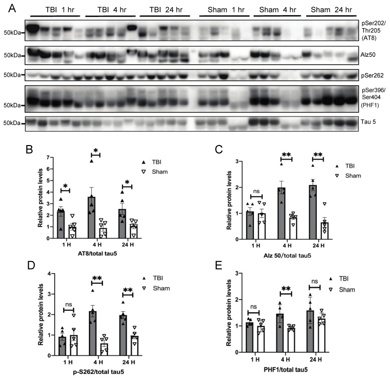
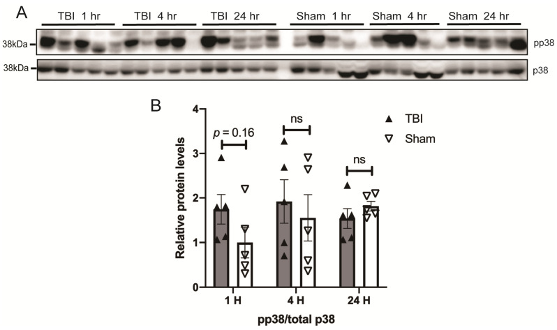
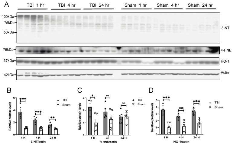
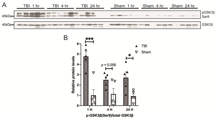
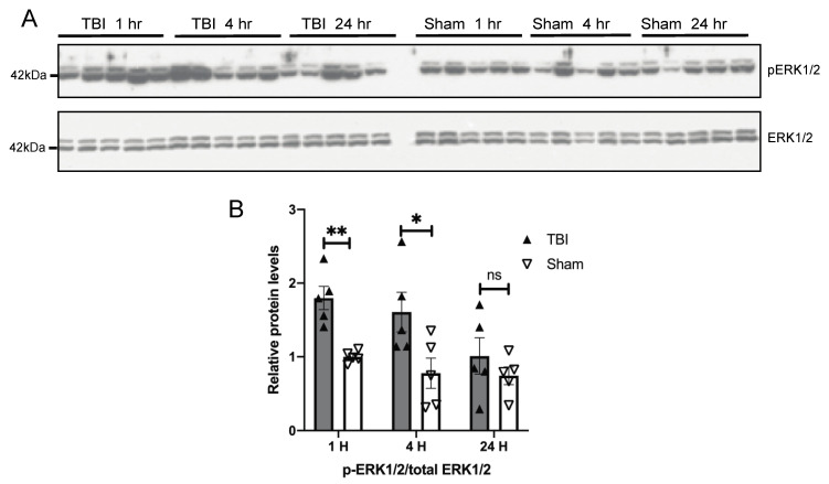
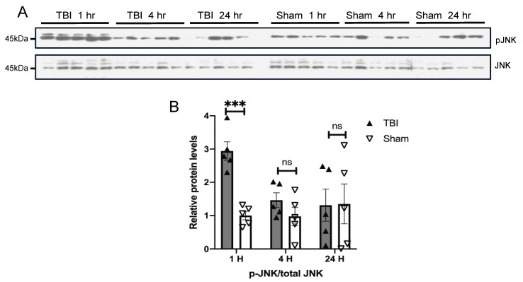
Traumatic brain injury caused by blast is associated with long-term neuropathological changes including tau phosphorylation and pathology. In this study, we aimed to determine changes in initial tau phosphorylation after exposure to a single mild blast and the potential contribution of oxidative stress response pathways. C57BL/6 mice were exposed to a single blast overpressure (BOP) generated by a compressed gas-driven shock tube that recapitulates battlefield-relevant open-field BOP, and cortical tissues were harvested at different time points up to 24 h after blast for Western blot analysis. We found that BOP caused elevated tau phosphorylation at Ser202/Thr205 detected by the AT8 antibody at 1 h post-blast followed by tau phosphorylation at additional sites (Ser262 and Ser396/Ser404 detected by PHF1 antibody) and conformational changes detected by Alz50 antibody. BOP also induced acute oxidative damage at 1 h post-blast and gradually declined overtime. Interestingly, Extracellular signal-regulated kinase (ERK) and c-Jun N-terminal kinase (JNK) were acutely activated in a similar temporal pattern as the rise and fall in oxidative stress after blast, with p38 showing a similar trend. However, glycogen synthase kinase-3 β (GSK3β) was inhibited at 1 h and remained inhibited for 24 h post blast. These results suggested that mitogen-activated protein kinases (MAPKs but not GSK3β are likely involved in mediating the effects of oxidative stress on the initial increase of tau phosphorylation following a single mild blast.
爆炸所致创伤性脑损伤与包括tau蛋白磷酸化和病理变化在内的长期神经病理学改变有关。在本研究中,我们旨在确定单次轻度爆炸暴露后初始tau蛋白磷酸化的变化以及氧化应激反应途径的潜在作用。将C57BL/6小鼠暴露于由压缩气体驱动的激波管产生的单次爆炸超压(BOP)下,该激波管模拟与战场相关的开阔场地BOP,在爆炸后长达24小时的不同时间点采集皮质组织进行蛋白质印迹分析。我们发现,爆炸后1小时,AT8抗体检测到BOP导致Ser202/Thr205位点的tau蛋白磷酸化升高,随后其他位点(PHF1抗体检测到的Ser262和Ser396/Ser404)的tau蛋白磷酸化以及Alz50抗体检测到的构象变化。BOP还在爆炸后1小时诱导急性氧化损伤,并随时间逐渐下降。有趣的是,细胞外信号调节激酶(ERK)和c-Jun氨基末端激酶(JNK)在爆炸后氧化应激的上升和下降过程中以类似的时间模式被急性激活,p38也呈现类似趋势。然而,糖原合酶激酶-3β(GSK3β)在爆炸后1小时被抑制,并在爆炸后24小时持续被抑制。这些结果表明,丝裂原活化蛋白激酶(MAPKs而非GSK3β)可能参与介导氧化应激对单次轻度爆炸后tau蛋白磷酸化初始增加的影响。