Division of Anatomical Pathology, Vancouver General Hospital, Vancouver, British Columbia, Canada.
Department of Pathology & Laboratory Medicine, University of British Columbia, Vancouver, British Columbia, Canada.
BMC Cancer. 2017 Sep 5;17(1):618. doi: 10.1186/s12885-017-3634-5.
Programmed cell death 1 (PD1) inhibitors have recently shown promising anti-cancer effects in a number of solid tumor types. A predictive biomarker to this class of drugs has not been clearly identified; however, overexpression of the PD1 ligand (PD-L1) has shown particular promise in lung adenocarcinoma. In this study, we explore the staining characteristics, prevalence, and clinico-molecular correlates of PD-L1 overexpression in pancreatic ductal adenocarcinoma (PDAC).
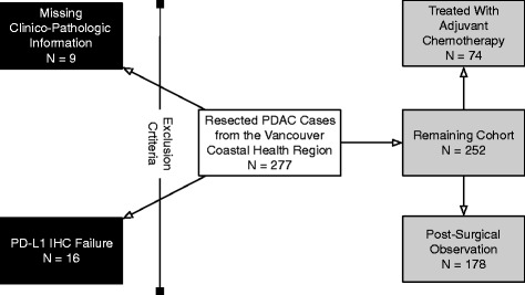
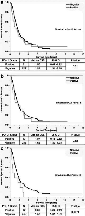
A tissue microarray (TMA) was constructed from cases of resected PDAC. PD-L1 immunohistochemistry (IHC) was performed using the SP142 primary antibody. Immunohistochemical assessment for deficient mismatch repair status (MMRd), CD3 and CD8 were performed. All biomarkers were assessed independently by two anatomical pathologists and consensus achieved on all cases. Survival analysis was performed using three thresholds (> = 1%, >5% and >10%) for tumor cell membrane staining.
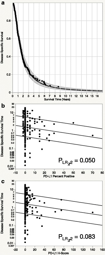
Two-hundred fifty-two cases were included in the TMA and evaluable by IHC. Thirty-one (12%), 17 (7%), 12(5%) cases were positive at percentage cut offs of >0, >5, and >10% respectively. Increased PD-L1 expression was associated with inferior prognosis (p = 0.0367). No statistically significant association was identified between PD-L1 status and MMR status or tumor infiltrating lymphocytes.
This data suggests that there is an inverse relationship between PD-L1 expression and disease specific survival times in resected PDAC. Consequently, this association may represent a phenotype where increased PD-L1 expression has an effect on tumor biology and could therefore identify a subgroup where PD1 blockade could have enhanced effectiveness.
程序性死亡受体 1(PD1)抑制剂在多种实体肿瘤类型中表现出了有前景的抗癌作用。但是,尚未明确此类药物的预测性生物标志物;然而,PD1 配体(PD-L1)的过表达在肺腺癌中显示出了特别的前景。在这项研究中,我们探讨了胰腺导管腺癌(PDAC)中 PD-L1 过表达的染色特征、流行率和临床分子相关性。
从切除的 PDAC 病例中构建组织微阵列(TMA)。使用 SP142 一抗进行 PD-L1 免疫组织化学(IHC)染色。进行免疫组织化学评估以确定错配修复缺陷状态(MMRd)、CD3 和 CD8。两位解剖病理学家独立评估所有生物标志物,并在所有病例上达成共识。使用肿瘤细胞膜染色的三个阈值(≥1%、>5%和>10%)进行生存分析。
在 TMA 中纳入了 252 例可进行 IHC 评估的病例。分别有 31(12%)、17(7%)和 12(5%)例病例在>0%、>5%和>10%的百分比截断值下为阳性。PD-L1 表达增加与预后不良相关(p=0.0367)。PD-L1 状态与 MMR 状态或肿瘤浸润淋巴细胞之间未发现统计学显著相关性。
该数据表明,在切除的 PDAC 中,PD-L1 表达与疾病特异性生存时间呈负相关。因此,这种相关性可能代表了一种表型,其中 PD-L1 表达增加对肿瘤生物学有影响,因此可以识别出 PD1 阻断可能增强疗效的亚组。