Jeong Ii-Do, Kim Woong-Chul, Park Jinyoung, Kim Chong-Myeong, Kim Ji-Hwan
Institute for Health Science, Korea University, Seoul, Republic of Korea.
Department of Dental Laboratory Science and Engineering, Korea University, Seoul, Republic of Korea.
J Adv Prosthodont. 2017 Aug;9(4):252-256. doi: 10.4047/jap.2017.9.4.252. Epub 2017 Aug 16.
This study aimed to analyze and compare the reproducibility of zirconia and lithium disilicate crowns manufactured by digital workflow.
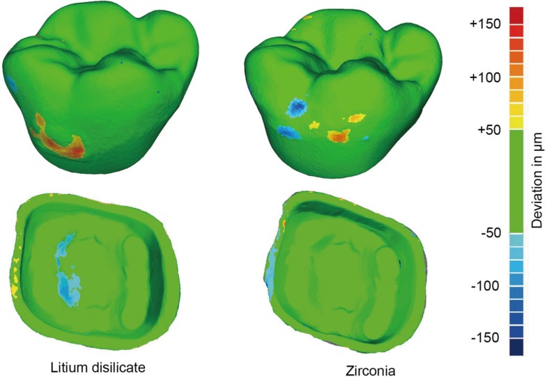
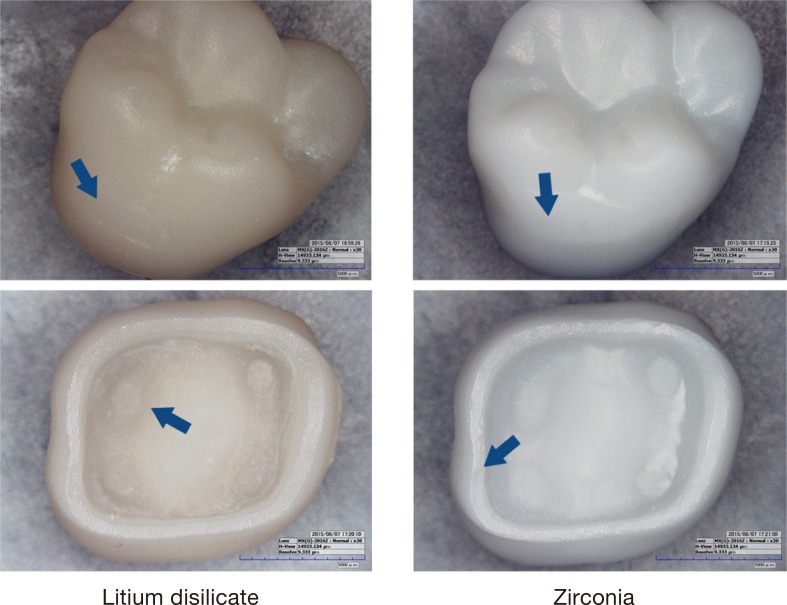
A typodont model with a prepped upper first molar was set in a phantom head, and a digital impression was obtained with a video intraoral scanner (CEREC Omnicam; Sirona GmbH), from which a single crown was designed and manufactured with CAD/CAM into a zirconia crown and lithium disilicate crown (n=12). Reproducibility of each crown was quantitatively retrieved by superimposing the digitized data of the crown in 3D inspection software, and differences were graphically mapped in color. Areas with large differences were analyzed with digital microscopy. Mean quadratic deviations (RMS) quantitatively obtained from each ceramic group were statistically analyzed with Student's t-test (α=.05).
The RMS value of lithium disilicate crown was 29.2 (4.1) µm and 17.6 (5.5) µm on the outer and inner surfaces, respectively, whereas these values were 18.6 (2.0) µm and 20.6 (5.1) µm for the zirconia crown. Reproducibility of zirconia and lithium disilicate crowns had a statistically significant difference only on the outer surface (<.001). The outer surface of lithium disilicate crown showed over-contouring on the buccal surface and under-contouring on the inner occlusal surface. The outer surface of zirconia crown showed both over- and under-contouring on the buccal surface, and the inner surface showed under-contouring in the marginal areas.
Restoration manufacturing by digital workflow will enhance the reproducibility of zirconia single crowns more than that of lithium disilicate single crowns.
本研究旨在分析和比较通过数字工作流程制造的氧化锆和二硅酸锂全冠的可重复性。
将带有预备好的上颌第一磨牙的牙列模型置于仿头模中,使用口腔内视频扫描仪(CEREC Omnicam;西诺德有限公司)获取数字印模,从中设计并使用CAD/CAM制造单个全冠,制成氧化锆全冠和二硅酸锂全冠(n = 12)。通过在三维检查软件中叠加全冠的数字化数据定量获取每个全冠的可重复性,并以颜色图形化映射差异。使用数字显微镜分析差异较大的区域。对每个陶瓷组定量获得的平均二次偏差(RMS)进行Student t检验统计分析(α = 0.05)。
二硅酸锂全冠外表面和内表面的RMS值分别为29.2(4.1)μm和17.6(5.5)μm,而氧化锆全冠的这些值分别为18.6(2.0)μm和20.6(5.1)μm。氧化锆和二硅酸锂全冠的可重复性仅在外表面有统计学显著差异(<.001)。二硅酸锂全冠的外表面在颊侧表面显示轮廓过度,在内侧咬合面显示轮廓不足。氧化锆全冠的外表面在颊侧表面显示轮廓过度和不足,内表面在边缘区域显示轮廓不足。
通过数字工作流程进行修复体制造,氧化锆单冠的可重复性比二硅酸锂单冠提高得更多。