van Leijsen Esther M C, van Uden Ingeborg W M, Ghafoorian Mohsen, Bergkamp Mayra I, Lohner Valerie, Kooijmans Eline C M, van der Holst Helena M, Tuladhar Anil M, Norris David G, van Dijk Ewoud J, Rutten-Jacobs Loes C A, Platel Bram, Klijn Catharina J M, de Leeuw Frank-Erik
From the Donders Institute for Brain, Cognition and Behaviour, Centre for Cognitive Neuroscience, Department of Neurology (E.M.C.v.L., I.W.M.v.U., M.I.B., V.L., E.C.M.K., H.M.v.d.H., A.M.T., E.J.v.D., C.J.M.K., F.-E.d.L.), and Diagnostic Image Analysis Group, Department of Radiology and Nuclear Medicine (M.G., B.P.), Radboud University Medical Centre; Institute for Computing and Information Sciences (M.G.) and Donders Institute for Brain, Cognition and Behaviour, Centre for Cognitive Neuroimaging (D.G.N.), Radboud University, Nijmegen, the Netherlands; Department of Clinical Neurosciences, Neurology Unit (L.C.A.R.-J.), University of Cambridge, UK; and Erwin L. Hahn Institute for Magnetic Resonance Imaging (D.G.N.), University of Duisburg-Essen, Essen, Germany.
Neurology. 2017 Oct 10;89(15):1569-1577. doi: 10.1212/WNL.0000000000004490. Epub 2017 Sep 6.
To investigate the temporal dynamics of cerebral small vessel disease (SVD) by 3 consecutive assessments over a period of 9 years, distinguishing progression from regression.
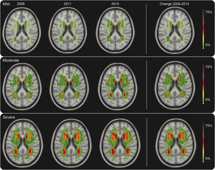
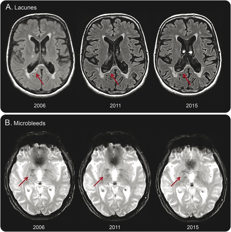
Changes in SVD markers of 276 participants of the Radboud University Nijmegen Diffusion Tensor and Magnetic Resonance Imaging Cohort (RUN DMC) cohort were assessed at 3 time points over 9 years. We assessed white matter hyperintensities (WMH) volume by semiautomatic segmentation and rated lacunes and microbleeds manually. We categorized baseline WMH severity as mild, moderate, or severe according to the modified Fazekas scale. We performed mixed-effects regression analysis including a quadratic term for increasing age.
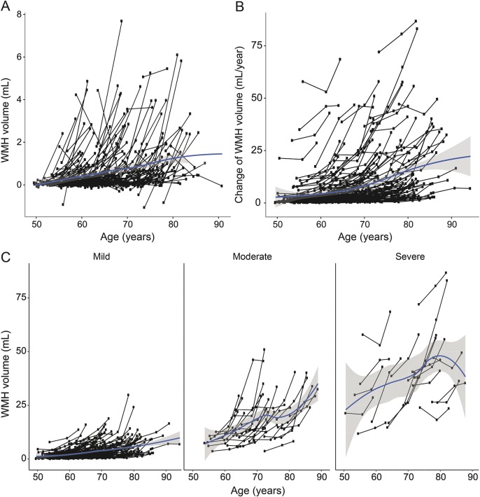
Mean WMH progression over 9 years was 4.7 mL (0.54 mL/y; interquartile range 0.95-5.5 mL), 20.3% of patients had incident lacunes (2.3%/y), and 18.9% had incident microbleeds (2.2%/y). WMH volume declined in 9.4% of the participants during the first follow-up interval, but only for 1 participant (0.4%) throughout the whole follow-up. Lacunes disappeared in 3.6% and microbleeds in 5.7% of the participants. WMH progression accelerated over time: including a quadratic term for increasing age during follow-up significantly improved the model ( < 0.001). SVD progression was predominantly seen in participants with moderate to severe WMH at baseline compared to those with mild WMH (odds ratio [OR] 35.5, 95% confidence interval [CI] 15.8-80.0, < 0.001 for WMH progression; OR 5.7, 95% CI 2.8-11.2, < 0.001 for incident lacunes; and OR 2.9, 95% CI 1.4-5.9, = 0.003 for incident microbleeds).
SVD progression is nonlinear, accelerating over time, and a highly dynamic process, with progression interrupted by reduction in some, in a population that on average shows progression.
通过在9年时间内进行3次连续评估,研究脑小血管病(SVD)的时间动态变化,区分病情进展与病情缓解。
对拉德堡大学奈梅亨分校弥散张量与磁共振成像队列(RUN DMC)中的276名参与者的SVD标志物变化在9年中的3个时间点进行了评估。我们通过半自动分割评估白质高信号(WMH)体积,并手动对腔隙和微出血进行评分。根据改良的 Fazekas 量表,我们将基线WMH严重程度分为轻度、中度或重度。我们进行了混合效应回归分析,包括年龄增长的二次项。
9年期间WMH的平均进展为4.7 mL(0.54 mL/年;四分位间距为0.95 - 5.5 mL),20.3%的患者出现新发腔隙(2.3%/年),18.9%的患者出现新发微出血(2.2%/年)。在第一次随访期间,9.4%的参与者WMH体积下降,但在整个随访过程中只有1名参与者(0.4%)出现这种情况。3.6%的参与者腔隙消失,5.7%的参与者微出血消失。WMH进展随时间加速:在随访期间纳入年龄增长的二次项显著改善了模型(P < 0.001)。与轻度WMH参与者相比,基线时中度至重度WMH参与者的SVD进展更为明显(WMH进展的优势比[OR]为35.5,95%置信区间[CI]为15.8 - 80.0,P < 0.001;新发腔隙的OR为5.7,95%CI为2.8 - (此处原文有误,应为11.2),P < 0.001;新发微出血的OR为2.9,95%CI为1.4 - 5.9,P = 0.003)。
SVD进展是非线性的,随时间加速,是一个高度动态的过程,在平均显示病情进展的人群中,部分病情进展会被病情缓解所打断。