• 文献检索
  • 文档翻译
  • 深度研究
  • 学术资讯
  • Suppr Zotero 插件Zotero 插件
  • 邀请有礼
  • 套餐&价格
  • 历史记录
应用&插件
Suppr Zotero 插件Zotero 插件浏览器插件Mac 客户端Windows 客户端微信小程序
定价
高级版会员购买积分包购买API积分包
服务
文献检索文档翻译深度研究API 文档MCP 服务
关于我们
关于 Suppr公司介绍联系我们用户协议隐私条款
关注我们

Suppr 超能文献

核心技术专利:CN118964589B侵权必究
粤ICP备2023148730 号-1Suppr @ 2026

文献检索

告别复杂PubMed语法,用中文像聊天一样搜索,搜遍4000万医学文献。AI智能推荐,让科研检索更轻松。

立即免费搜索

文件翻译

保留排版,准确专业,支持PDF/Word/PPT等文件格式,支持 12+语言互译。

免费翻译文档

深度研究

AI帮你快速写综述,25分钟生成高质量综述,智能提取关键信息,辅助科研写作。

立即免费体验

在认知正常的老年人中,脑白质高信号与皮质厚度之间存在相互增强关系的纵向证据。

Longitudinal evidence for a mutually reinforcing relationship between white matter hyperintensities and cortical thickness in cognitively unimpaired older adults.

机构信息

Institute of Cognitive Neurology and Dementia Research, Otto-Von-Guericke University Magdeburg, Magdeburg, Germany.

German Centre for Neurodegenerative Diseases (DZNE), Magdeburg, Germany.

出版信息

Alzheimers Res Ther. 2024 Oct 28;16(1):240. doi: 10.1186/s13195-024-01606-5.

DOI:10.1186/s13195-024-01606-5
PMID:39465440
原文链接:https://pmc.ncbi.nlm.nih.gov/articles/PMC11520063/
Abstract

BACKGROUND

For over three decades, the concomitance of cortical neurodegeneration and white matter hyperintensities (WMH) has sparked discussions about their coupled temporal dynamics. Longitudinal studies supporting this hypothesis nonetheless remain scarce.

METHODS

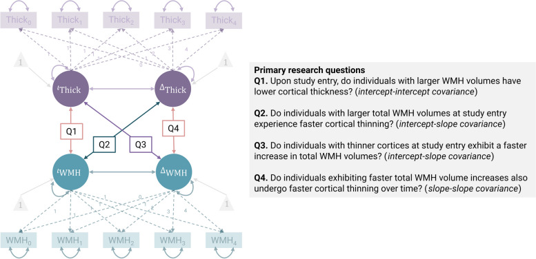
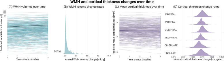
We applied global and regional bivariate latent growth curve modelling to determine the extent to which WMH and cortical thickness were interrelated over a four-year period. For this purpose, we leveraged longitudinal MRI data from 451 cognitively unimpaired participants (DELCODE; median age 69.71 [IQR 65.51, 75.50] years; 52.32% female). Participants underwent MRI sessions annually over a four-year period (1815 sessions in total, with roughly four MRI sessions per participant). We adjusted all models for demographics and cardiovascular risk.

RESULTS

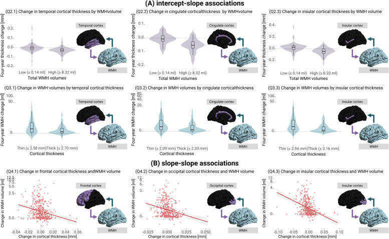
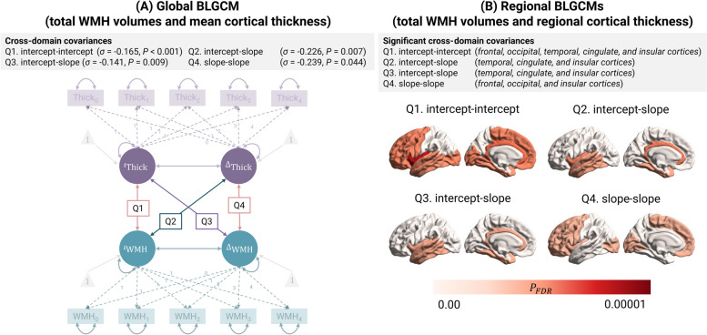
Our findings were three-fold. First, larger WMH volumes were linked to lower cortical thickness (σ = -0.165, SE = 0.047, Z = -3.515, P < 0.001). Second, individuals with higher WMH volumes experienced more rapid cortical thinning (σ = -0.226, SE = 0.093, Z = -2.443, P = 0.007), particularly in temporal, cingulate, and insular regions. Similarly, those with lower initial cortical thickness had faster WMH progression (σ = -0.141, SE = 0.060, Z = -2.336, P = 0.009), with this effect being most pronounced in temporal, cingulate, and insular cortices. Third, faster WMH progression was associated with accelerated cortical thinning (σ = -0.239, SE = 0.139, Z = -1.710, P = 0.044), particularly in frontal, occipital, and insular cortical regions.

CONCLUSIONS

Our study suggests that cortical thinning and WMH progression could be mutually reinforcing rather than parallel, unrelated processes, which become entangled before cognitive deficits are detectable.

TRIAL REGISTRATION

German Clinical Trials Register (DRKS00007966, 04/05/2015).

摘要

背景

三十多年来,皮质神经退行性变和脑白质高信号(WMH)的同时存在引发了关于它们时间动态耦合的讨论。然而,支持这一假设的纵向研究仍然很少。

方法

我们应用全局和区域双变量潜增长曲线模型来确定 WMH 和皮质厚度在四年期间相互关联的程度。为此,我们利用来自 451 名认知正常的参与者的纵向 MRI 数据(DELCODE;中位年龄 69.71 [IQR 65.51, 75.50] 岁;52.32%女性)。参与者在四年期间每年接受 MRI 检查(共 1815 次,每位参与者约有四次 MRI 检查)。我们调整了所有模型以适应人口统计学和心血管风险因素。

结果

我们的研究结果有三个方面。首先,较大的 WMH 体积与较低的皮质厚度相关(σ=-0.165,SE=0.047,Z=-3.515,P<0.001)。其次,WMH 体积较高的个体经历了更快的皮质变薄(σ=-0.226,SE=0.093,Z=-2.443,P=0.007),特别是在颞叶、扣带回和岛叶区域。同样,初始皮质厚度较低的个体有更快的 WMH 进展(σ=-0.141,SE=0.060,Z=-2.336,P=0.009),这种效应在颞叶、扣带回和岛叶皮质最为明显。第三,WMH 进展较快与皮质变薄加速相关(σ=-0.239,SE=0.139,Z=-1.710,P=0.044),特别是在额叶、枕叶和岛叶皮质区域。

结论

我们的研究表明,皮质变薄和 WMH 进展可能是相互促进的,而不是平行的、不相关的过程,在可检测到认知缺陷之前,这些过程就已经纠缠在一起了。

试验注册

德国临床试验注册处(DRKS00007966,2015 年 4 月 5 日)。

https://cdn.ncbi.nlm.nih.gov/pmc/blobs/ab8b/11520063/f5e8afe120d2/13195_2024_1606_Fig4_HTML.jpg
https://cdn.ncbi.nlm.nih.gov/pmc/blobs/ab8b/11520063/5994e56891c4/13195_2024_1606_Fig1_HTML.jpg
https://cdn.ncbi.nlm.nih.gov/pmc/blobs/ab8b/11520063/6f7acdbd37f8/13195_2024_1606_Fig2_HTML.jpg
https://cdn.ncbi.nlm.nih.gov/pmc/blobs/ab8b/11520063/efeade2243b9/13195_2024_1606_Fig3_HTML.jpg
https://cdn.ncbi.nlm.nih.gov/pmc/blobs/ab8b/11520063/f5e8afe120d2/13195_2024_1606_Fig4_HTML.jpg

相似文献

1
Longitudinal evidence for a mutually reinforcing relationship between white matter hyperintensities and cortical thickness in cognitively unimpaired older adults.在认知正常的老年人中,脑白质高信号与皮质厚度之间存在相互增强关系的纵向证据。
Alzheimers Res Ther. 2024 Oct 28;16(1):240. doi: 10.1186/s13195-024-01606-5.
2
Association of Regional White Matter Hyperintensities With Longitudinal Alzheimer-Like Pattern of Neurodegeneration in Older Adults.老年人大脑中与阿尔茨海默病样神经退行性变相关的区域性脑白质高信号与纵向变化的关联。
JAMA Netw Open. 2021 Oct 1;4(10):e2125166. doi: 10.1001/jamanetworkopen.2021.25166.
3
Linking white matter hyperintensities to regional cortical thinning, amyloid deposition, and synaptic density loss in Alzheimer's disease.将脑白质高信号与阿尔茨海默病的区域性皮质变薄、淀粉样蛋白沉积和突触密度丧失联系起来。
Alzheimers Dement. 2024 Jun;20(6):3931-3942. doi: 10.1002/alz.13845. Epub 2024 Apr 22.
4
Clinical and Biological Correlates of White Matter Hyperintensities in Patients With Behavioral-Variant Frontotemporal Dementia and Alzheimer Disease.行为变异型额颞叶痴呆和阿尔茨海默病患者脑白质高信号的临床和生物学相关性。
Neurology. 2021 Mar 30;96(13):e1743-e1754. doi: 10.1212/WNL.0000000000011638. Epub 2021 Feb 17.
5
Progression of White Matter Disease and Cortical Thinning Are Not Related in Older Community-Dwelling Subjects.在社区居住的老年人中,白质病变进展与皮质变薄无关。
Stroke. 2016 Feb;47(2):410-6. doi: 10.1161/STROKEAHA.115.011229. Epub 2015 Dec 22.
6
The relationship between cortical thickness and white matter hyperintensities in mid to late life.中年至老年期皮质厚度与白质高信号之间的关系。
Neurobiol Aging. 2024 Sep;141:129-139. doi: 10.1016/j.neurobiolaging.2024.05.014. Epub 2024 May 29.
7
Regional cortical thinning, demyelination and iron loss in cerebral small vessel disease.脑小血管病的区域性皮质变薄、脱髓鞘和铁丢失。
Brain. 2023 Nov 2;146(11):4659-4673. doi: 10.1093/brain/awad220.
8
Rates of cortical thinning in Alzheimer's disease signature regions associate with vascular burden but not with β-amyloid status in cognitively normal adults at age 70.在 70 岁认知正常的成年人中,阿尔茨海默病特征区域的皮质变薄率与血管负担相关,而与β-淀粉样蛋白状态无关。
J Neurol Neurosurg Psychiatry. 2024 Jul 15;95(8):748-752. doi: 10.1136/jnnp-2023-332067.
9
The effect of white matter hyperintensities on cognition is mediated by cortical atrophy.脑白质高信号对认知的影响是通过皮质萎缩介导的。
Neurobiol Aging. 2018 Apr;64:25-32. doi: 10.1016/j.neurobiolaging.2017.12.006. Epub 2017 Dec 16.
10
Associations between white matter hyperintensities, lacunes, entorhinal cortex thickness, declarative memory and leisure activity in cognitively healthy older adults: A 7-year study.认知健康老年人脑白质高信号、腔隙、内嗅皮层厚度、陈述性记忆和休闲活动之间的关联:一项 7 年研究。
Neuroimage. 2023 Dec 15;284:120461. doi: 10.1016/j.neuroimage.2023.120461. Epub 2023 Nov 17.

引用本文的文献

1
Brain Microstructural Alteration and Its Implications for Postoperative Delirium in Elderly Surgical Patients: A Narrative Review.脑微结构改变及其对老年外科手术患者术后谵妄的影响:一项叙述性综述
Brain Behav. 2025 Sep;15(9):e70872. doi: 10.1002/brb3.70872.
2
Recent advances in neuroimaging of Alzheimer's disease and related dementias.阿尔茨海默病及相关痴呆症神经影像学的最新进展。
Alzheimers Dement. 2025 Sep;21(9):e70648. doi: 10.1002/alz.70648.
3
White matter hyperintensities and their impact in brain structure and function in alzheimer's disease and behavioral variant frontotemporal dementia across Latin America and the United States: a cross-sectional study.

本文引用的文献

1
Interplay of physical and recognition performance using hierarchical continuous-time dynamic modeling and a dual-task training regime in Alzheimer's patients.在阿尔茨海默病患者中,使用分层连续时间动态建模和双任务训练方案对身体和认知表现的相互作用进行研究。
Alzheimers Dement (Amst). 2024 Aug 26;16(3):e12629. doi: 10.1002/dad2.12629. eCollection 2024 Jul-Sep.
2
CAT: a computational anatomy toolbox for the analysis of structural MRI data.CAT:用于分析结构磁共振成像数据的计算解剖工具箱。
Gigascience. 2024 Jan 2;13. doi: 10.1093/gigascience/giae049.
3
LST-AI: A deep learning ensemble for accurate MS lesion segmentation.
白质高信号及其对拉丁美洲和美国阿尔茨海默病及行为变异型额颞叶痴呆脑结构和功能的影响:一项横断面研究。
Alzheimers Res Ther. 2025 Aug 12;17(1):188. doi: 10.1186/s13195-025-01832-5.
LST-AI:用于精确 MS 病变分割的深度学习集成。
Neuroimage Clin. 2024;42:103611. doi: 10.1016/j.nicl.2024.103611. Epub 2024 Apr 29.
4
Linking white matter hyperintensities to regional cortical thinning, amyloid deposition, and synaptic density loss in Alzheimer's disease.将脑白质高信号与阿尔茨海默病的区域性皮质变薄、淀粉样蛋白沉积和突触密度丧失联系起来。
Alzheimers Dement. 2024 Jun;20(6):3931-3942. doi: 10.1002/alz.13845. Epub 2024 Apr 22.
5
Associations between white matter hyperintensities, lacunes, entorhinal cortex thickness, declarative memory and leisure activity in cognitively healthy older adults: A 7-year study.认知健康老年人脑白质高信号、腔隙、内嗅皮层厚度、陈述性记忆和休闲活动之间的关联:一项 7 年研究。
Neuroimage. 2023 Dec 15;284:120461. doi: 10.1016/j.neuroimage.2023.120461. Epub 2023 Nov 17.
6
Etiology of White Matter Hyperintensities in Autosomal Dominant and Sporadic Alzheimer Disease.常染色体显性遗传和散发性阿尔茨海默病患者脑白质高信号的病因。
JAMA Neurol. 2023 Dec 1;80(12):1353-1363. doi: 10.1001/jamaneurol.2023.3618.
7
New horizons in cognitive and functional impairment as a consequence of cerebral small vessel disease.脑小血管病导致认知和功能障碍的新领域。
Age Ageing. 2023 Aug 1;52(8). doi: 10.1093/ageing/afad148.
8
Regional cortical thinning, demyelination and iron loss in cerebral small vessel disease.脑小血管病的区域性皮质变薄、脱髓鞘和铁丢失。
Brain. 2023 Nov 2;146(11):4659-4673. doi: 10.1093/brain/awad220.
9
Neuroimaging standards for research into small vessel disease-advances since 2013.神经影像学在小血管疾病研究中的标准——2013 年以来的进展。
Lancet Neurol. 2023 Jul;22(7):602-618. doi: 10.1016/S1474-4422(23)00131-X. Epub 2023 May 23.
10
Arterial hypertension and β-amyloid accumulation have spatially overlapping effects on posterior white matter hyperintensity volume: a cross-sectional study.动脉高血压和β-淀粉样蛋白堆积对后部脑白质高信号体积具有空间上重叠的影响:一项横断面研究。
Alzheimers Res Ther. 2023 May 24;15(1):97. doi: 10.1186/s13195-023-01243-4.