• 文献检索
  • 文档翻译
  • 深度研究
  • 学术资讯
  • Suppr Zotero 插件Zotero 插件
  • 邀请有礼
  • 套餐&价格
  • 历史记录
应用&插件
Suppr Zotero 插件Zotero 插件浏览器插件Mac 客户端Windows 客户端微信小程序
定价
高级版会员购买积分包购买API积分包
服务
文献检索文档翻译深度研究API 文档MCP 服务
关于我们
关于 Suppr公司介绍联系我们用户协议隐私条款
关注我们

Suppr 超能文献

核心技术专利:CN118964589B侵权必究
粤ICP备2023148730 号-1Suppr @ 2026

文献检索

告别复杂PubMed语法,用中文像聊天一样搜索,搜遍4000万医学文献。AI智能推荐,让科研检索更轻松。

立即免费搜索

文件翻译

保留排版,准确专业,支持PDF/Word/PPT等文件格式,支持 12+语言互译。

免费翻译文档

深度研究

AI帮你快速写综述,25分钟生成高质量综述,智能提取关键信息,辅助科研写作。

立即免费体验

移植物抗宿主病相关干眼的活体共聚焦显微镜评估。

In Vivo Confocal Microscopy Evaluation of Ocular Surface with Graft-Versus-Host Disease-Related Dry Eye Disease.

机构信息

Aier School of Ophthalmology, Central South University, Changsha, China.

Department of Ophthalmology, Keio University School of Medicine, Tokyo, Japan.

出版信息

Sci Rep. 2017 Sep 6;7(1):10720. doi: 10.1038/s41598-017-10237-w.

DOI:10.1038/s41598-017-10237-w
PMID:28878217
原文链接:https://pmc.ncbi.nlm.nih.gov/articles/PMC5587759/
Abstract

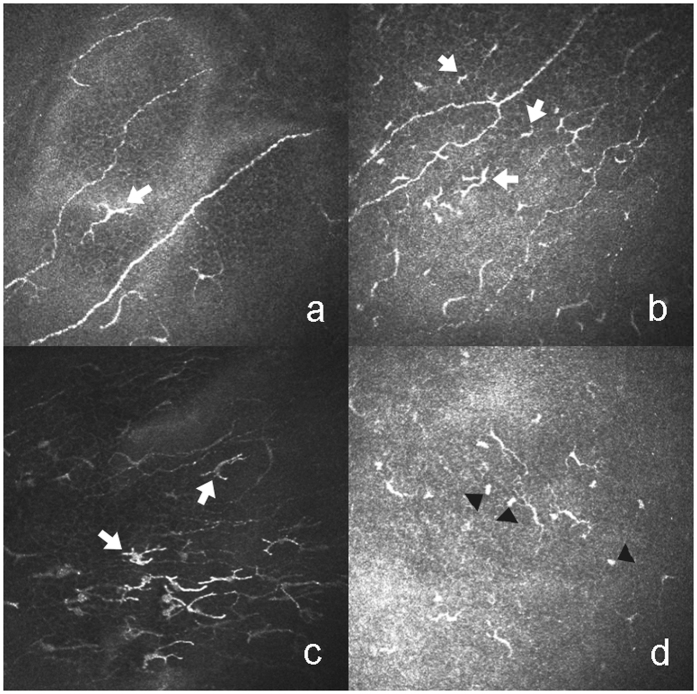
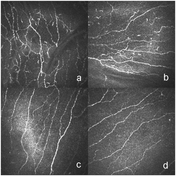
Dry eye disease (DED) is often elicited by graft-versus-host disease (GVHD), an extensive complication of hematopoietic stem cell transplantation (HSCT). To unravel the mechanism of this type of DED, in vivo confocal microscopy (IVCM) was used to investigate alterations in the state of the sub-basal nerves, dendritic cells (DCs) and globular immune cells (GICs) in the central cornea and limbal epithelia. In this study, we examined 12 HSCT recipients with GVHD-caused DED and 10 HSCT recipients without GVHD-associated DED and evaluated the clinical parameters in the 2 groups. Analysis of the central cornea and limbal epithelia using IVCM was conducted to investigate the density of the corneal sub-basal nerves, DCs and GICs as well as the tortuosity and branching of the sub-basal nerves. As suggested by our data, the clinical variables in the GVHD group were significantly different from those in the non-GVHD group. Additionally, GVHD-triggered DED conceivably increased the density of DCs and GICs in the central cornea and the density of DCs in limbal epithelia and altered the morphology of the sub-basal nerves. These phenomena are presumably correlated with the degree of inflammation. Thus, our findings may be translated into non-invasive diagnostic methods that indicate the severity of inflammation on the ocular surface in HSCT recipients.

摘要

干眼症(DED)通常由移植物抗宿主病(GVHD)引起,GVHD 是造血干细胞移植(HSCT)的一种广泛并发症。为了阐明这种类型的 DED 的机制,我们使用共聚焦显微镜(IVCM)研究了中央角膜和角膜缘上皮的基底下神经、树突状细胞(DC)和球状免疫细胞(GIC)状态的变化。在这项研究中,我们检查了 12 例因 GVHD 引起的 DED 的 HSCT 受者和 10 例无 GVHD 相关 DED 的 HSCT 受者,并评估了两组的临床参数。通过 IVCM 对中央角膜和角膜缘上皮进行分析,以研究角膜基底下神经、DC 和 GIC 的密度以及基底下神经的迂曲和分支。正如我们的数据所表明的,GVHD 组的临床变量与非 GVHD 组明显不同。此外,GVHD 引发的 DED 可能会增加中央角膜中 DC 和 GIC 的密度以及角膜缘上皮中 DC 的密度,并改变基底下神经的形态。这些现象可能与炎症程度有关。因此,我们的发现可以转化为非侵入性诊断方法,这些方法可以指示 HSCT 受者眼表面的炎症严重程度。

https://cdn.ncbi.nlm.nih.gov/pmc/blobs/6243/5587759/a3e2f9e76233/41598_2017_10237_Fig3_HTML.jpg
https://cdn.ncbi.nlm.nih.gov/pmc/blobs/6243/5587759/ebd3ac7d50e0/41598_2017_10237_Fig1_HTML.jpg
https://cdn.ncbi.nlm.nih.gov/pmc/blobs/6243/5587759/c5c6e645c151/41598_2017_10237_Fig2_HTML.jpg
https://cdn.ncbi.nlm.nih.gov/pmc/blobs/6243/5587759/a3e2f9e76233/41598_2017_10237_Fig3_HTML.jpg
https://cdn.ncbi.nlm.nih.gov/pmc/blobs/6243/5587759/ebd3ac7d50e0/41598_2017_10237_Fig1_HTML.jpg
https://cdn.ncbi.nlm.nih.gov/pmc/blobs/6243/5587759/c5c6e645c151/41598_2017_10237_Fig2_HTML.jpg
https://cdn.ncbi.nlm.nih.gov/pmc/blobs/6243/5587759/a3e2f9e76233/41598_2017_10237_Fig3_HTML.jpg

相似文献

1
In Vivo Confocal Microscopy Evaluation of Ocular Surface with Graft-Versus-Host Disease-Related Dry Eye Disease.移植物抗宿主病相关干眼的活体共聚焦显微镜评估。
Sci Rep. 2017 Sep 6;7(1):10720. doi: 10.1038/s41598-017-10237-w.
2
In Vivo Confocal Microscopy in Dry Eye Disease Associated With Chronic Graft-Versus-Host Disease.与慢性移植物抗宿主病相关的干眼病的体内共聚焦显微镜检查
Invest Ophthalmol Vis Sci. 2016 Sep 1;57(11):4686-91. doi: 10.1167/iovs.16-20013.
3
Corneal features in ocular graft-versus-host disease by in vivo confocal microscopy.活体共聚焦显微镜观察眼移植物抗宿主病的角膜特征
Graefes Arch Clin Exp Ophthalmol. 2017 Dec;255(12):2389-2397. doi: 10.1007/s00417-017-3759-x. Epub 2017 Sep 5.
4
In vivo confocal microscopic characterisation of the cornea in chronic graft-versus-host disease related severe dry eye disease.慢性移植物抗宿主病相关严重干眼病中角膜的体内共聚焦显微镜特征分析
Br J Ophthalmol. 2015 Feb;99(2):160-5. doi: 10.1136/bjophthalmol-2014-305072. Epub 2014 Aug 19.
5
In vivo confocal microscopy morphometric analysis of corneal subbasal nerve plexus in dry eye disease using newly developed fully automated system.使用新开发的全自动系统对干眼症患者角膜基底神经丛进行体内共焦显微镜形态计量分析。
Graefes Arch Clin Exp Ophthalmol. 2019 Mar;257(3):583-589. doi: 10.1007/s00417-018-04225-7. Epub 2019 Jan 14.
6
Corneal Epithelial Immune Dendritic Cell Alterations in Subtypes of Dry Eye Disease: A Pilot In Vivo Confocal Microscopic Study.干眼疾病亚型中角膜上皮免疫树突状细胞的改变:一项体内共聚焦显微镜初步研究
Invest Ophthalmol Vis Sci. 2015 Nov;56(12):7179-85. doi: 10.1167/iovs.15-17433.
7
[Clinical characteristics and correlative factors of dry eye disease associated with graft-versus-host disease].移植物抗宿主病相关干眼的临床特征及相关因素
Zhonghua Yan Ke Za Zhi. 2023 Apr 11;59(4):295-301. doi: 10.3760/cma.j.cn112142-20220801-00370.
8
Evaluation of aqueous-deficient and evaporative dry eye cases with confocal microscopy.应用共焦显微镜评估水液缺乏性和蒸发过强型干眼症。
J Fr Ophtalmol. 2023 Dec;46(10):1161-1168. doi: 10.1016/j.jfo.2023.05.024. Epub 2023 Sep 25.
9
How does ocular graft-versus-host disease fit under the dry eye umbrella? A review.眼部移植物抗宿主病如何归入干眼范畴?一篇综述。
Clin Exp Ophthalmol. 2024 Mar;52(2):167-185. doi: 10.1111/ceo.14347. Epub 2024 Jan 10.
10
Can in vivo confocal microscopy differentiate between sub-types of dry eye disease? A review.活体共聚焦显微镜能否区分干眼疾病的亚型?综述。
Clin Exp Ophthalmol. 2021 May;49(4):373-387. doi: 10.1111/ceo.13924. Epub 2021 Apr 13.

引用本文的文献

1
Translating Biomarker Discovery: From Bench to Bedside in Dry Eye Disease.干眼症生物标志物发现的转化:从实验室到临床应用
Int J Mol Sci. 2025 Sep 3;26(17):8556. doi: 10.3390/ijms26178556.
2
[Ocular graft-versus-host disease : S1 guidelines of the German Society of Ophthalmology (DOG) and the German Professional Association of Ophthalmologists (BVA)].[眼部移植物抗宿主病:德国眼科学会(DOG)和德国眼科医生专业协会(BVA)的S1指南]
Ophthalmologie. 2025 Sep 1. doi: 10.1007/s00347-025-02296-6.
3
In vivo Confocal Microscopy for Corneal and Ocular Surface Pathologies: A Comprehensive Review.

本文引用的文献

1
Heavy Chain-Hyaluronan/Pentraxin 3 from Amniotic Membrane Suppresses Inflammation and Scarring in Murine Lacrimal Gland and Conjunctiva of Chronic Graft-versus-Host Disease.羊膜来源重链-透明质酸/五聚素 3 抑制慢性移植物抗宿主病小鼠泪腺和结膜的炎症和瘢痕形成。
Sci Rep. 2017 Feb 6;7:42195. doi: 10.1038/srep42195.
2
Novel Treatment of Chronic Graft-Versus-Host Disease in Mice Using the ER Stress Reducer 4-Phenylbutyric Acid.使用内质网应激抑制剂 4-苯丁酸治疗小鼠慢性移植物抗宿主病的新方法。
Sci Rep. 2017 Feb 6;7:41939. doi: 10.1038/srep41939.
3
In Vivo Distribution of Corneal Epithelial Dendritic Cells in Patients With Glaucoma.
用于角膜和眼表疾病的体内共聚焦显微镜检查:全面综述。
Clin Ophthalmol. 2025 Jun 7;19:1817-1834. doi: 10.2147/OPTH.S519705. eCollection 2025.
4
Ophthalmic Manifestations in Patients with Blood Malignancies.血液系统恶性肿瘤患者的眼部表现
Hematol Rep. 2024 Mar 28;16(2):193-203. doi: 10.3390/hematolrep16020020.
5
Advances in the ocular complications after hematopoietic stem cell transplantation.造血干细胞移植后眼部并发症的研究进展。
Ann Hematol. 2024 Oct;103(10):3867-3880. doi: 10.1007/s00277-024-05678-z. Epub 2024 Feb 26.
6
Development and validation of a diagnostic model for the identification of chronic ocular graft-versus-host disease (oGVHD).用于识别慢性眼部移植物抗宿主病(oGVHD)的诊断模型的开发与验证
Front Med (Lausanne). 2023 Oct 27;10:1277194. doi: 10.3389/fmed.2023.1277194. eCollection 2023.
7
[Chinese expert consensus on ocular chronic graft-versus-host disease (2023)].《中国眼部慢性移植物抗宿主病专家共识(2023年)》
Zhonghua Xue Ye Xue Za Zhi. 2023 Jun 14;44(6):449-457. doi: 10.3760/cma.j.issn.0253-2727.2023.06.002.
8
Challenges and concepts in the diagnosis and management of ocular graft-versus-host disease.眼部移植物抗宿主病诊断与管理中的挑战与概念
Front Med (Lausanne). 2023 Feb 16;10:1133381. doi: 10.3389/fmed.2023.1133381. eCollection 2023.
9
The ocular graft-versus-host disease: the path from current knowledge to future managements.眼移植物抗宿主病:从现有知识到未来管理的途径。
Eye (Lond). 2023 Jul;37(10):1982-1992. doi: 10.1038/s41433-022-02288-9. Epub 2022 Nov 4.
10
A novel combination of corneal confocal microscopy, clinical features and artificial intelligence for evaluation of ocular surface pain.新型角膜共焦显微镜、临床特征和人工智能联合评估眼表疼痛。
PLoS One. 2022 Nov 1;17(11):e0277086. doi: 10.1371/journal.pone.0277086. eCollection 2022.
青光眼患者角膜上皮树突状细胞的体内分布
Invest Ophthalmol Vis Sci. 2016 Nov 1;57(14):5996-6002. doi: 10.1167/iovs.16-20333.
4
In Vivo Confocal Microscopy in Dry Eye Disease Associated With Chronic Graft-Versus-Host Disease.与慢性移植物抗宿主病相关的干眼病的体内共聚焦显微镜检查
Invest Ophthalmol Vis Sci. 2016 Sep 1;57(11):4686-91. doi: 10.1167/iovs.16-20013.
5
In vivo confocal microscopy evaluation of ocular and cutaneous alterations in patients with rosacea.酒渣鼻患者眼部和皮肤改变的体内共聚焦显微镜评估
Br J Ophthalmol. 2017 Mar;101(3):268-274. doi: 10.1136/bjophthalmol-2015-308110. Epub 2016 May 24.
6
Corneal Epithelial Immune Dendritic Cell Alterations in Subtypes of Dry Eye Disease: A Pilot In Vivo Confocal Microscopic Study.干眼疾病亚型中角膜上皮免疫树突状细胞的改变:一项体内共聚焦显微镜初步研究
Invest Ophthalmol Vis Sci. 2015 Nov;56(12):7179-85. doi: 10.1167/iovs.15-17433.
7
Contralateral Clinically Unaffected Eyes of Patients With Unilateral Infectious Keratitis Demonstrate a Sympathetic Immune Response.单侧感染性角膜炎患者对侧临床未受累眼表现出交感免疫反应。
Invest Ophthalmol Vis Sci. 2015 Oct;56(11):6612-20. doi: 10.1167/iovs.15-16560.
8
Degeneration and Regeneration of Subbasal Corneal Nerves after Infectious Keratitis: A Longitudinal In Vivo Confocal Microscopy Study.感染性角膜炎后角膜基底膜下神经的退变与再生:一项纵向活体共聚焦显微镜研究
Ophthalmology. 2015 Nov;122(11):2200-9. doi: 10.1016/j.ophtha.2015.06.047. Epub 2015 Aug 6.
9
Vision-Related Quality of Life in Patients with Ocular Graft-versus-Host Disease.眼部移植物抗宿主病患者与视觉相关的生活质量
Ophthalmology. 2015 Aug;122(8):1669-74. doi: 10.1016/j.ophtha.2015.04.011. Epub 2015 May 20.
10
Dry eye and corneal langerhans cells in systemic lupus erythematosus.系统性红斑狼疮中的干眼与角膜朗格汉斯细胞
J Ophthalmol. 2015;2015:543835. doi: 10.1155/2015/543835. Epub 2015 Mar 29.