Varadarajan Indumathy, Basu Aparna, Besmer Sherri, Poli Jaganmohan, Richard Scott, Styler Michael
Drexel University College of Medicine, Philadelphia, Pennsylvania, USA.
Henry Ford Medical Center, Detroit, Michigan, USA.
Case Rep Oncol. 2017 Aug 4;10(2):694-698. doi: 10.1159/000478976. eCollection 2017 May-Aug.
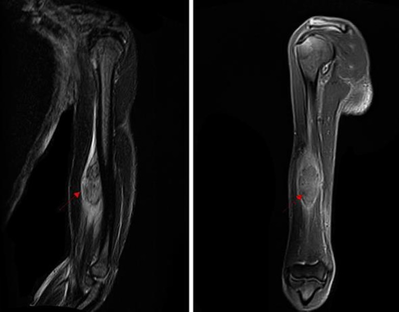
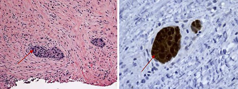
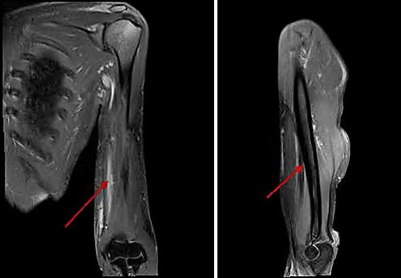
Cervical cancer is the fourth most common cancer in women worldwide, with a large majority of prevalence (85%) in developing countries. As of 2012, it accounts for 7.5% of all female cancer deaths. Despite its high prevalence, skeletal muscle metastasis from cervical cancer is extremely uncommon. In our extensive literature search, we were able to find only 8 cases where skeletal muscle metastasis was the only site of recurrence. We report a case of a 52-year-old African-American woman with a past medical history of cervical cancer (stage IIIB) who presented with pain and swelling in her left upper arm over the preceding 2 months. MRI of the left upper arm showed a solid well-circumscribed mass measuring 7.0 × 2.8 × 2.5 cm, deep to the biceps. Biopsy of the mass revealed a metastatic squamous cell carcinoma that was p16-positive. PET scan showed that the lesion was the sole site of metastasis. She received local radiation with concurrent chemotherapy. Follow-up MRI 6 months after the completion of therapy showed resolution of the mass. She has remained disease-free for the last 24 months as evidenced by a PET/CT scan in May 2016. In this case report, we discuss the role of imaging and pathology in the diagnosis of a solitary metastatic lesion. This case also emphasizes the importance of a close follow-up which aids in early intervention, increasing overall survival.
宫颈癌是全球女性中第四大常见癌症,在发展中国家的患病率占绝大多数(85%)。截至2012年,宫颈癌占所有女性癌症死亡人数的7.5%。尽管其患病率很高,但宫颈癌的骨骼肌转移极为罕见。在我们广泛的文献检索中,我们仅能找到8例骨骼肌转移是唯一复发部位的病例。我们报告一例52岁非裔美国女性,既往有宫颈癌病史(IIIB期),在过去2个月出现左上臂疼痛和肿胀。左上臂MRI显示一个边界清晰的实性肿块,大小为7.0×2.8×2.5cm,位于肱二头肌深部。肿块活检显示为p16阳性的转移性鳞状细胞癌。PET扫描显示该病变是唯一的转移部位。她接受了局部放疗并同时进行化疗。治疗结束6个月后的随访MRI显示肿块消退。2016年5月的PET/CT扫描显示,在过去24个月里她一直无病生存。在本病例报告中,我们讨论了影像学和病理学在孤立性转移病变诊断中的作用。本病例还强调了密切随访的重要性,这有助于早期干预,提高总体生存率。