Department of Anesthesiology and Critical Care Medicine, David Geffen School of Medicine, University of California, Los AngelesLos Angeles, CA, United States.
Front Neural Circuits. 2017 Aug 25;11:58. doi: 10.3389/fncir.2017.00058. eCollection 2017.
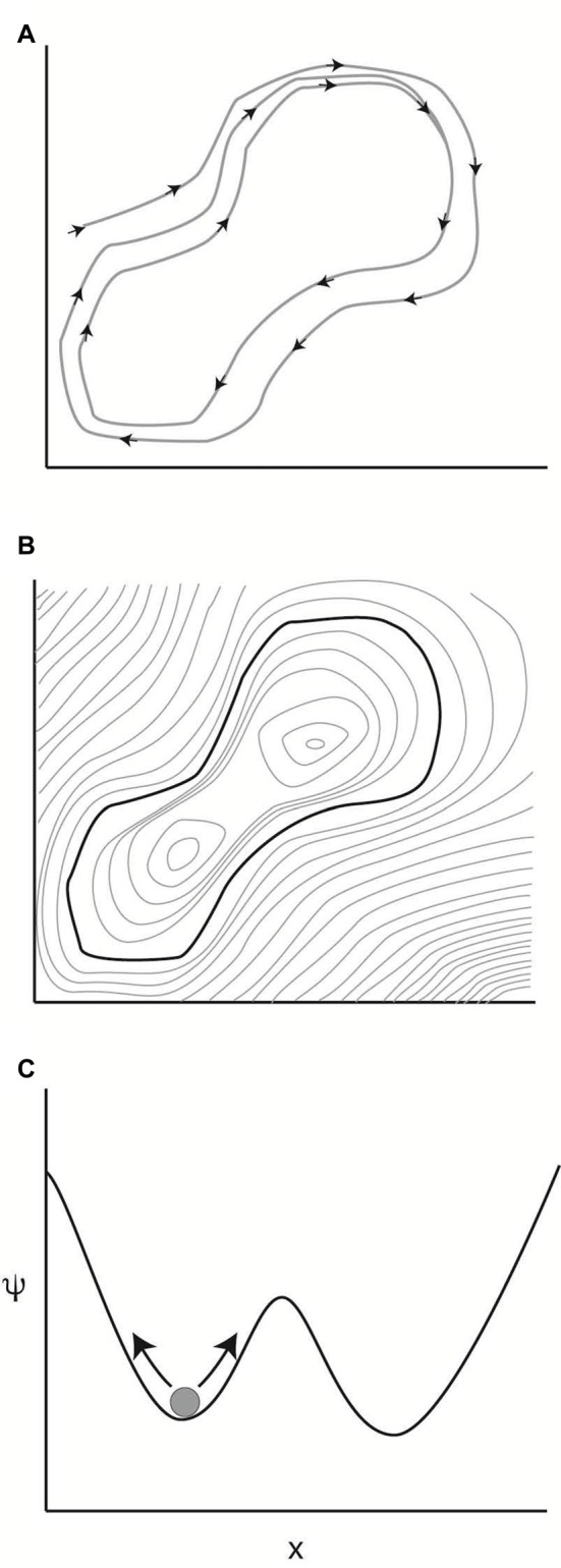
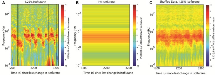
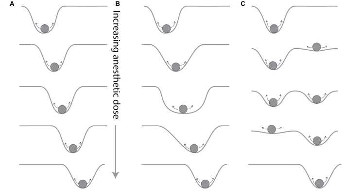
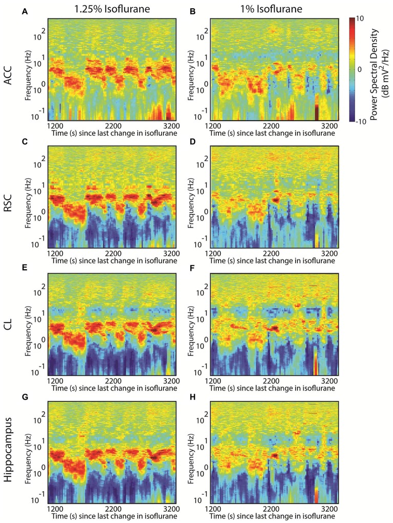
There is strong evidence that anesthetics have stereotypical effects on brain state, so that a given anesthetic appears to have a signature in the electroencephalogram (EEG), which may vary with dose. This can be usefully interpreted as the anesthetic determining an attractor in the phase space of the brain. How brain activity shifts between these attractors in time remains understudied, as most studies implicitly assume a one-to-one relationship between drug dose and attractor features by assuming stationarity over the analysis interval and analyzing data segments of several minutes in length. Yet data in rats anesthetized with isoflurane suggests that, at anesthetic levels consistent with surgical anesthesia, brain activity alternates between multiple attractors, often spending on the order of 10 min in one activity pattern before shifting to another. Moreover, the probability of these jumps between attractors changes with anesthetic concentration. This suggests the hypothesis that brain state is metastable during anesthesia: though it appears at equilibrium on short timescales (on the order of seconds to a few minutes), longer intervals show shifting behavior. Compelling evidence for metastability in rats anesthetized with isoflurane is reviewed, but so far only suggestive hints of metastability in brain states exist with other anesthetics or in other species. Explicit testing of metastability during anesthesia will require experiments with longer acquisition intervals and carefully designed analytic approaches; some of the implications of these constraints are reviewed for typical spectral analysis approaches. If metastability exists during anesthesia, it implies degeneracy in the relationship between brain state and effect site concentration, as there is not a one-to-one mapping between the two. This degeneracy could explain some of the reported difficulty in using brain activity monitors to titrate drug dose to prevent awareness during anesthesia and should force a rethinking of the notion of depth of anesthesia as a single dimension. Finally, explicit incorporation of knowledge of the dynamics of the brain during anesthesia could offer better depth of anesthesia monitoring.
有强有力的证据表明,麻醉剂对大脑状态有刻板的影响,因此,给定的麻醉剂在脑电图(EEG)中似乎具有特征,其特征可能随剂量而变化。这可以被有效地解释为麻醉剂确定大脑相空间中的吸引子。大脑活动在这些吸引子之间如何随时间转移仍在研究中,因为大多数研究通过在分析间隔内假设平稳性并分析几分钟长的数据段,从而隐含地假设药物剂量和吸引子特征之间存在一一对应关系。然而,在异氟烷麻醉的大鼠中获得的数据表明,在与手术麻醉一致的麻醉水平下,大脑活动在多个吸引子之间交替,通常在一种活动模式中花费大约 10 分钟的时间,然后转移到另一种活动模式。此外,这些吸引子之间跳跃的概率随麻醉剂浓度而变化。这表明了一个假设,即在麻醉期间大脑状态是亚稳态的:尽管它在短时间尺度(几秒钟到几分钟)上看起来处于平衡状态,但较长的间隔显示出转移行为。在异氟烷麻醉的大鼠中,对亚稳性的有力证据进行了综述,但到目前为止,只有其他麻醉剂或其他物种中存在脑状态亚稳性的提示性迹象。在麻醉期间进行亚稳性的明确测试将需要具有更长采集间隔和精心设计的分析方法的实验;对于典型的谱分析方法,回顾了这些约束的一些含义。如果在麻醉期间存在亚稳性,则意味着大脑状态与效应部位浓度之间的关系存在退化,因为两者之间没有一一对应的映射。这种退化可以解释一些报告的在使用脑活动监测仪来滴定药物剂量以防止麻醉期间意识的困难,并且应该迫使重新思考麻醉深度作为单一维度的概念。最后,在麻醉期间明确纳入对大脑动力学的知识可以提供更好的麻醉深度监测。