Ruzycki Philip A, Linne Courtney D, Hennig Anne K, Chen Shiming
Molecular Genetics and Genomics Graduate Program, Division of Biology & Biomedical Sciences, Washington University, St. Louis, Missouri, United States.
Department of Ophthalmology and Visual Sciences, Washington University, St. Louis, Missouri, United States.
Invest Ophthalmol Vis Sci. 2017 Sep 1;58(11):4644-4653. doi: 10.1167/iovs.17-22075.
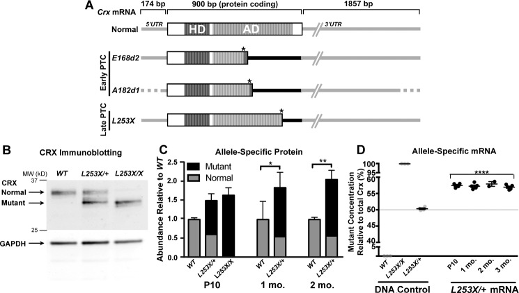
The cone-rod homeobox (CRX) transcription factor is essential for photoreceptor gene expression, differentiation, and survival. Human CRX mutations can cause dominant retinopathies of varying onset and phenotype severity. In animal models, dominant frameshift Crx mutations introduce a premature termination codon (PTC), producing inactive truncated proteins that interfere with normal CRX function. Previously, a mutant mouse, TVRM65, was reported to carry a recessive late PTC mutation, Crx-L253X. More detailed phenotype analysis of the pathogenicity of Crx-L253X sheds new light on the variability of CRX-linked diseases.
Homozygous (L253X/X); heterozygous (L253X/+); Crx-/- and control C57BL/6J (WT) mice were analyzed at various ages for changes in retinal function (ERG), morphology (histology) and photoreceptor gene expression (qRT-PCR).
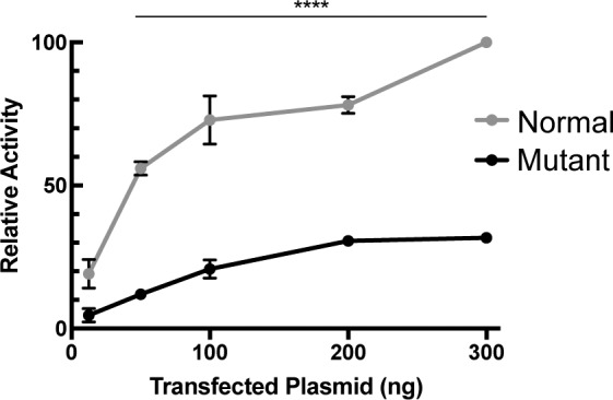
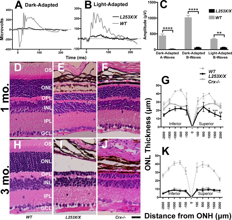
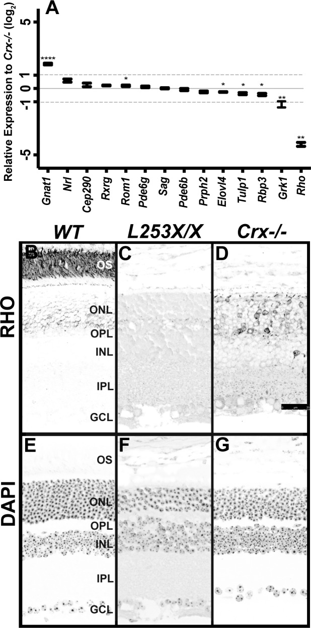
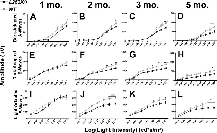
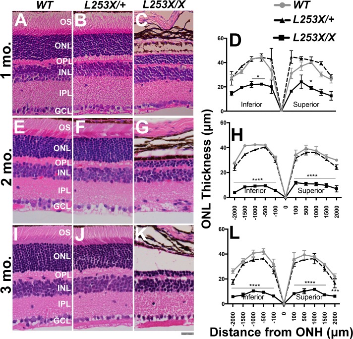
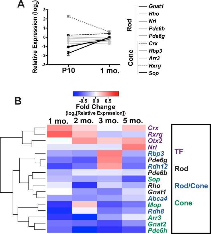
At 1 month, L253X/X mice lack visual function, show greater reductions in retinal thickness, and distinct gene expression changes relative to Crx-/-, suggesting that the phenotype of L253X/X is more severe than Crx-/-. L253X/+ mice have reduced rod/cone function, but normal retinal morphology at all ages tested. qRT-PCR assays described a complex phenotype in which both developing and mature photoreceptors are unable to maintain proper gene expression. L253X mRNA/protein is overexpressed relative to normal Crx, suggesting a pathogenic mechanism similar to early PTC mutations. However, the overexpression is less pronounced, correlating with a relatively mild dominant phenotype.
The L253X mouse provides a valuable model for CRX-associated retinopathy. The pathogenicity of CRX frameshift mutations depends on the position of the PTC, which in turn determines the degree of mutant mRNA/protein overproduction.
视锥-视杆同源框(CRX)转录因子对于光感受器基因表达、分化及存活至关重要。人类CRX突变可导致不同发病时间和表型严重程度的显性视网膜病变。在动物模型中,显性移码Crx突变引入过早终止密码子(PTC),产生无活性的截短蛋白,干扰正常CRX功能。此前,有报道称突变小鼠TVRM65携带隐性晚期PTC突变Crx-L253X。对Crx-L253X致病性进行更详细的表型分析,为CRX相关疾病的变异性提供了新线索。
对纯合子(L253X/X)、杂合子(L253X/+)、Crx基因敲除小鼠(Crx-/-)及对照C57BL/6J野生型(WT)小鼠在不同年龄进行视网膜功能(视网膜电图,ERG)、形态学(组织学)及光感受器基因表达(定量逆转录聚合酶链反应,qRT-PCR)变化分析。
1月龄时,L253X/X小鼠缺乏视觉功能,视网膜厚度降低幅度大于Crx-/-小鼠,且基因表达变化明显,提示L253X/X小鼠的表型比Crx-/-小鼠更严重。L253X/+小鼠在所有测试年龄的视杆/视锥功能均降低,但视网膜形态正常。qRT-PCR分析显示出一种复杂表型,即发育中和成熟的光感受器均无法维持正常基因表达。相对于正常Crx,L253X的mRNA/蛋白表达上调,提示其致病机制与早期PTC突变相似。然而,这种上调不太明显,与相对较轻的显性表型相关。
L253X小鼠为CRX相关视网膜病变提供了有价值的模型。CRX移码突变的致病性取决于PTC的位置,而这又决定了突变mRNA/蛋白的过量产生程度。