Niklaus Monique, Adams Olivia, Berezowska Sabina, Zlobec Inti, Graber Franziska, Slotta-Huspenina Julia, Nitsche Ulrich, Rosenberg Robert, Tschan Mario P, Langer Rupert
Institute of Pathology, University of Bern, CH-3008 Bern, Switzerland.
Graduate School for Cellular and Biomedical Sciences, University of Bern, CH-3008 Bern, Switzerland.
Oncotarget. 2017 May 2;8(33):54604-54615. doi: 10.18632/oncotarget.17554. eCollection 2017 Aug 15.
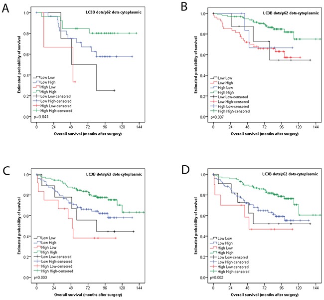
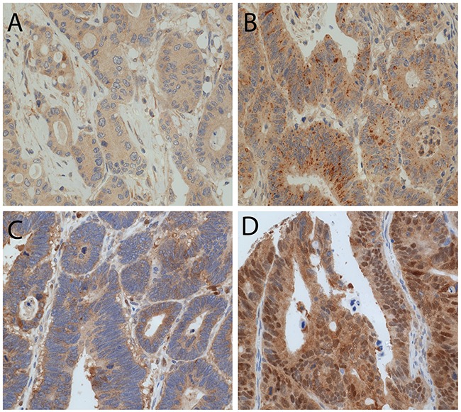
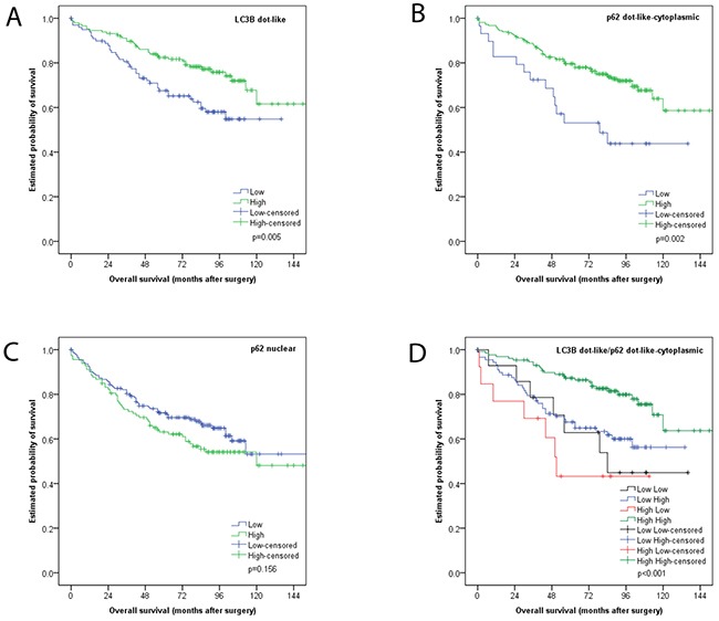
Autophagy is a lysosomal degradation and recycling process implicated in cancer progression and therapy resistance. We assessed the impact of basal autophagy in colon cancer (CC) and . Functional autophagy was demonstrated in CC cell lines (LoVo; HT-29) showing a dose-dependent increase of the autophagy markers LC3B, p62 and autophagic vesciles upon increasing concentrations of the autophagy inhibitor chloroquine, which was demonstrated by immunoblotting, immunofluorescence and electron microscopy. Next, tissue microarrays with 292 primary resected CC, with cores from different tumor regions, and normal mucosa were analyzed by immunohistochemistry for LC3B and p62. CC tissue showed LC3B dot-like, p62 dot-like, cytoplasmic and nuclear staining in various levels without significant intratumoral heterogeneity. Tumoral LC3B and p62 expression was significantly higher than in normal tissue (p<0.001). No associations between staining patterns and pathological features (e.g. TNM categories; grading) were observed. Both low LC3B dot-like and low p62 dot-like-cytoplasmic staining were associated with worse overall survival (p=0.005 and p=0.002). The best prognostic discrimination, however, was seen for a combination of LC3B dot-like/p62 dot-like-cytoplasmic staining: high expression of both markers, indicative of impaired activated autophagy, was associated with the best overall survival. In contrast, high LC3B dot-like/low p62 dot-like-cytoplasmic expression, indicative of intact activated autophagy, was associated with the worst outcome (p<0.001 in univariate and HR=0.751; CI=0.607-0.928; p=0.008 in multivariate analysis). These specific expression patterns of LC3B and p62 pointing to different states of autophagy associated with diverging clinical outcomes highlighte the potential significance of basal autophagy in CC biology.
自噬是一种溶酶体降解和再循环过程,与癌症进展和治疗耐药性有关。我们评估了基础自噬对结肠癌(CC)的影响。在CC细胞系(LoVo;HT-29)中证实了功能性自噬,随着自噬抑制剂氯喹浓度的增加,自噬标志物LC3B、p62和自噬囊泡呈剂量依赖性增加,这通过免疫印迹、免疫荧光和电子显微镜得以证实。接下来,通过免疫组织化学分析了包含292例原发性切除CC的组织芯片,这些组织芯片的核心来自不同肿瘤区域以及正常黏膜,用于检测LC3B和p62。CC组织显示出不同水平的LC3B点状、p62点状、细胞质和细胞核染色,肿瘤内无明显异质性。肿瘤组织中LC3B和p62的表达显著高于正常组织(p<0.001)。未观察到染色模式与病理特征(如TNM分类;分级)之间的关联。低LC3B点状染色和低p62点状细胞质染色均与较差的总生存期相关(p=0.005和p=0.002)。然而,对于LC3B点状/p62点状细胞质染色的组合,预后判别最佳:两种标志物的高表达表明激活自噬受损,与最佳总生存期相关。相反,高LC3B点状/低p62点状细胞质表达表明激活自噬完整,与最差预后相关(单变量分析中p<0.001,多变量分析中HR=0.751;CI=0.607-0.928;p=0.008)。LC3B和p62的这些特定表达模式指向与不同临床结果相关的自噬不同状态,突出了基础自噬在CC生物学中的潜在重要性。