Botolin Sergiu, VanderHeiden Todd F, Moore Ernest E, Fried Herbert, Stahel Philip F
Department of Orthopaedics, University of Colorado, School of Medicine and Denver Health Medical Center, 777 Bannock Street, Denver, CO 80204 USA.
Department of Surgery, University of Colorado, School of Medicine and Denver Health Medical Center, Denver, CO 80204 USA.
Patient Saf Surg. 2017 Sep 8;11:23. doi: 10.1186/s13037-017-0139-8. eCollection 2017.
Cervical spine fracture-dislocations in neurologically intact patients represent a surgical challenge due to the risk of inflicting iatrogenic spinal cord compression by closed reduction maneuvers. The use of MRI for early advanced imaging in these injuries remains controversially debated.
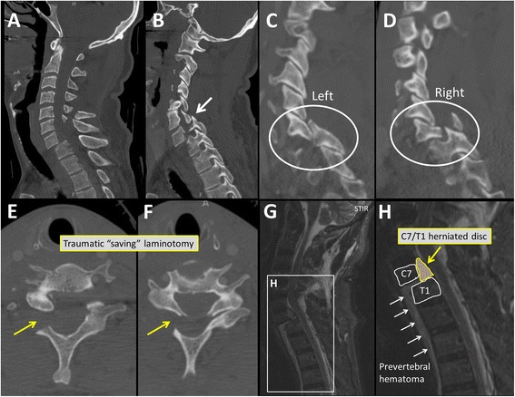
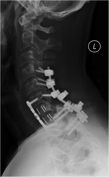
A 54-year old man sustained a fall over the handlebars of his racing bicycle. The helmeted patient sustained a fall on his head which resulted in a hyperflexion injury of the neck. He was neurologically intact on presentation. Initial CT imaging revealed a complex multisegmental cervical spine injury with a left-sided C6/C7 perched facet, a right sided C7/T1 fracture-dislocation, and a right-sided C6 and C7 traumatic laminotomy. The initial management consisted of temporary external Halo fixator application without closed reduction maneuver, to mitigate the risk of a delayed spinal cord injury. Subsequent advanced imaging by MRI revealed an acute traumatic C7/T1 disc herniation, with the intervertebral disc completely extruded into the spinal canal. Definitive surgical management was then accomplished by employing a three-stage anterior-posterior-anterior spinal decompression, realignment, fixation and fusion C4-T2 in one operative session. The patient recovered well and retained full neurological function. He resumed bicycle street racing within 10 months of the injury following successful spinal reconstruction.
The diagnostic evaluation of cervical fracture-dislocations should include advanced imaging by MRI in order to fully understand the injury pattern prior to proceeding with spinal reduction maneuvers which may impose the imminent threat of a devastating iatrogenic injury to the spinal cord. The presented staged management by initial Halo fixation without attempts for spinal reduction, followed by a surgical decompression and multilevel fusion, appears to represent a feasible and safe strategy for patients at risk of a delayed neurological injury.
对于神经功能完好的患者,颈椎骨折脱位是一项手术挑战,因为闭合复位操作有造成医源性脊髓压迫的风险。在这些损伤中使用MRI进行早期高级成像仍存在争议。
一名54岁男性从他的赛车车把上摔下。戴头盔的患者头部着地,导致颈部过屈损伤。就诊时他神经功能完好。初始CT成像显示复杂的多节段颈椎损伤,左侧C6/C7关节突交锁,右侧C7/T1骨折脱位,以及右侧C6和C7创伤性椎板切除术。初始治疗包括临时应用外部头环固定器,不进行闭合复位操作,以降低延迟性脊髓损伤的风险。随后通过MRI进行的高级成像显示急性创伤性C7/T1椎间盘突出,椎间盘完全突入椎管。然后通过在一次手术中采用前后前三级脊柱减压、复位、固定和融合C4-T2完成确定性手术治疗。患者恢复良好,保留了全部神经功能。在成功进行脊柱重建后,他在受伤后10个月内恢复了自行车公路赛。
颈椎骨折脱位的诊断评估应包括通过MRI进行高级成像,以便在进行可能对脊髓造成毁灭性医源性损伤的紧迫威胁的脊柱复位操作之前,充分了解损伤模式。所提出的初始采用头环固定而不尝试脊柱复位,随后进行手术减压和多节段融合的分期治疗方法,似乎是对有延迟性神经损伤风险的患者一种可行且安全的策略。