Gaddipati Vamsi C, Martin Angel I, Valenzuela Mauricio O, Mahmud Asef, Patel Aarti A
Department of Cardiovascular Sciences, Morsani College of Medicine, University of South Florida, Tampa, FL, USA.
Department of Internal Medicine, Morsani College of Medicine, University of South Florida, Tampa, FL, USA.
Case Rep Cardiol. 2017;2017:4352474. doi: 10.1155/2017/4352474. Epub 2017 Aug 24.
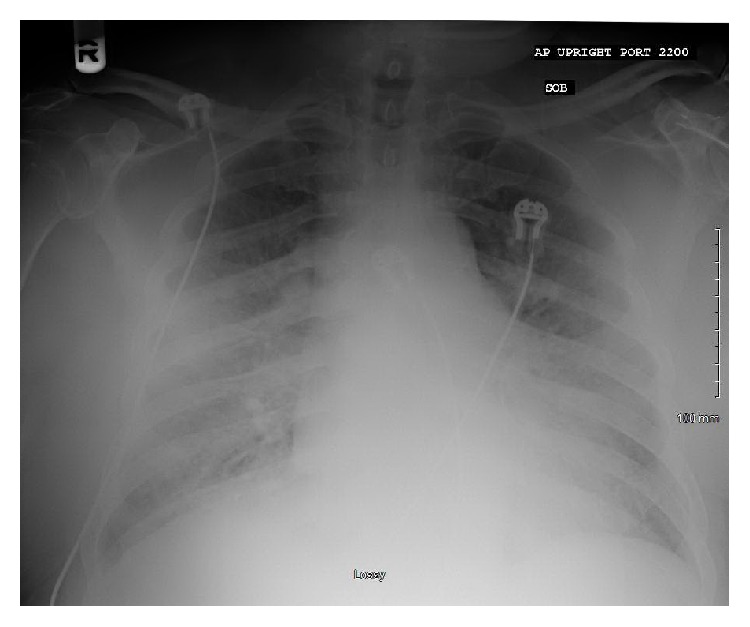
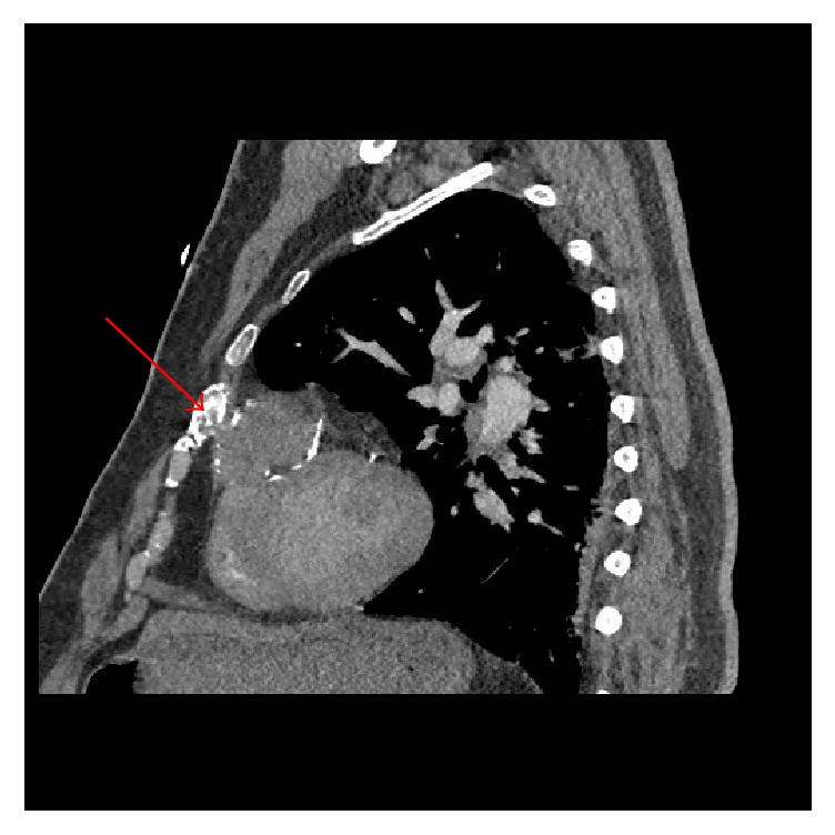
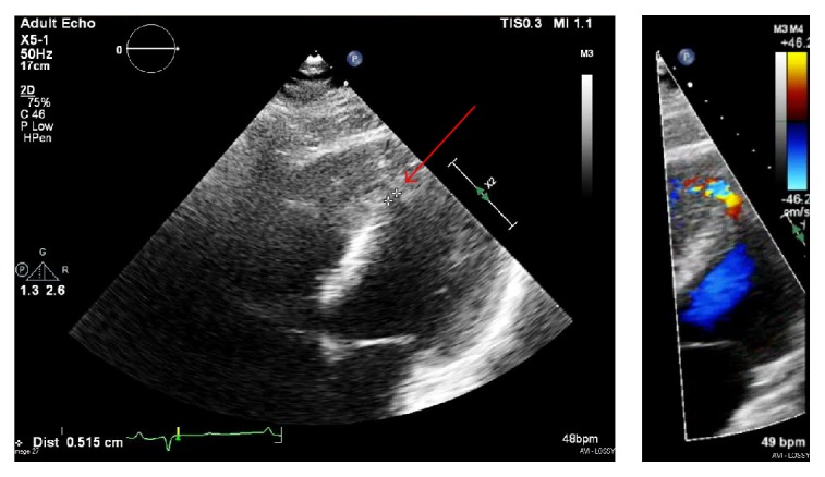
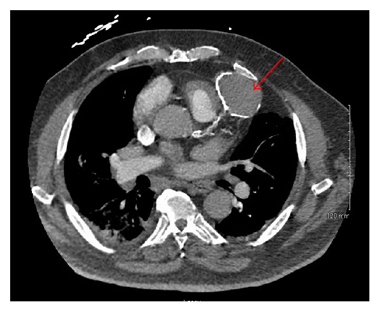
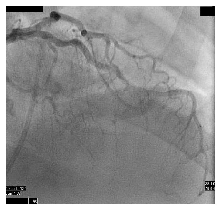
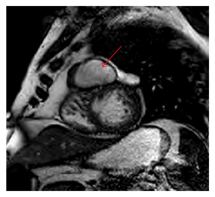
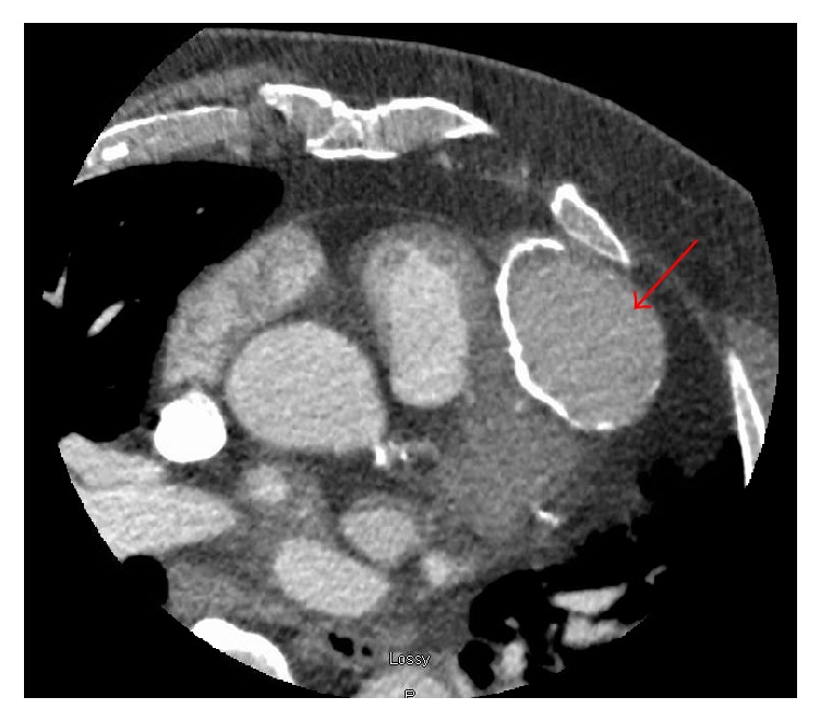
Ventricular pseudoaneurysm is an uncommon, potentially fatal complication that has been associated with myocardial infarction, cardiac surgery, chest trauma, and infectious processes. Diagnosis can be challenging, as cases are rare and slowly progressing and typically lack identifiable features on clinical presentation. As a result, advanced imaging techniques have become the hallmark of identification. Ahead, we describe a patient who presents with acute decompensated heart failure and was incidentally discovered to have a large right ventricular pseudoaneurysm that developed following previous traumatic anterior rib fracture.
心室假性动脉瘤是一种罕见的、潜在致命的并发症,与心肌梗死、心脏手术、胸部创伤及感染性疾病相关。诊断具有挑战性,因为病例罕见且进展缓慢,临床表现通常缺乏可识别特征。因此,先进的成像技术已成为识别的标志。在此,我们描述一名表现为急性失代偿性心力衰竭的患者,该患者在之前的外伤性前肋骨骨折后意外发现患有一个巨大的右心室假性动脉瘤。