Kim Ui Geon, Kook Dong Bee, Kim Tae Hun, Kim Chung Hun
Department of Plastic and Reconstructive Surgery, Bundang CHA Medical Center, CHA University School of Medicine, Seongnam, Korea.
Department of Pathology, Bundang CHA Medical Center, CHA University School of Medicine, Seongnam, Korea.
Arch Craniofac Surg. 2017 Mar;18(1):50-53. doi: 10.7181/acfs.2017.18.1.50. Epub 2017 Mar 25.
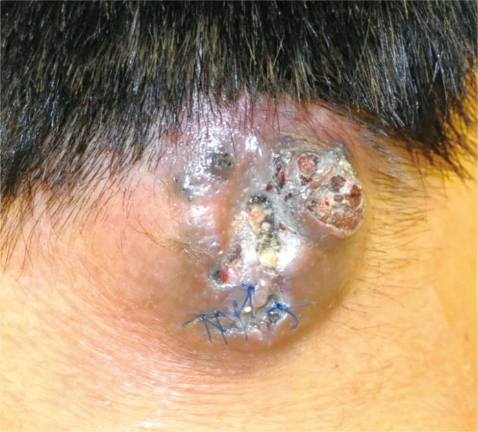
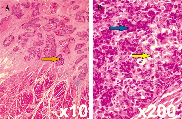
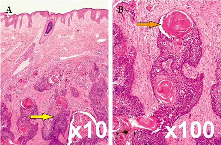
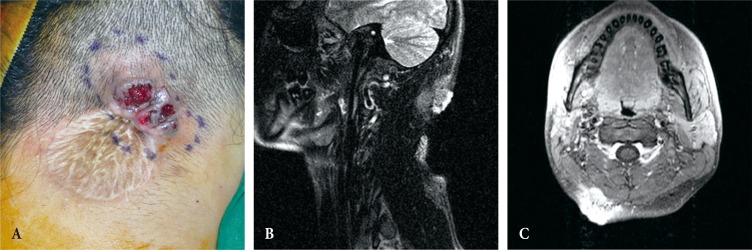
Trichilemmal cysts are common fluid-filled growths that arise from the isthmus of the hair follicle. They can form rapidly multiplying trichilemmal tumors-, also called proliferating trichilemmal cysts, which are typically benign. Rarely, proliferating trichilemmal cysts can become cancerous. Here we report the case of a patient who experienced this series of changes. The 27-year-old male patient had been observed to have a 1×1 cm cyst 7 years ago. Eight months prior to presentation at our institution, incision and drainage was performed at his local clinic. However, the size of the mass had gradually increased. At our clinic, he presented with a 5×4 cm hard mass that had recurred on the posterior side of his neck. The tumor was removed without safety margin, and the skin defect was covered with a split-thickness skin graft. The pathologic diagnosis was a benign proliferating trichilemmal cyst. The mass recurred after 4months, at which point, a wide excision (1.3-cm safety margin) and split-thickness skin graft were performed. The biopsy revealed a trichilemmal carcinoma arising from a proliferating trichilemmal cyst. This clinical experience suggests that clinicians should consider the possibility of malignant changes when diagnosing and treating trichilemmal cysts.
外毛根鞘囊肿是常见的充满液体的肿物,起源于毛囊峡部。它们可形成迅速增殖的外毛根鞘肿瘤,也称为增殖性外毛根鞘囊肿,通常为良性。增殖性外毛根鞘囊肿很少会发生癌变。在此,我们报告一例经历了这一系列变化的患者。该27岁男性患者7年前被发现有一个1×1 cm的囊肿。在他到我院就诊前8个月,其在当地诊所接受了切开引流。然而,肿物大小逐渐增大。在我院就诊时,他颈部后侧出现了一个复发的5×4 cm硬肿物。肿瘤在未留安全切缘的情况下被切除,皮肤缺损处用中厚皮片覆盖。病理诊断为良性增殖性外毛根鞘囊肿。4个月后肿物复发,此时进行了广泛切除(1.3 cm安全切缘)及中厚皮片移植。活检显示由增殖性外毛根鞘囊肿恶变而来的外毛根鞘癌。这一临床经验提示临床医生在诊断和治疗外毛根鞘囊肿时应考虑恶变的可能性。