Thuillier Philippe, Bourhis David, Robin Philippe, Keromnes Nathalie, Schick Ulrike, Le Roux Pierre-Yves, Kerlan Véronique, Chaumet-Riffaud Philippe, Salaün Pierre-Yves, Abgral Ronan
Department of Endocrinology, University Hospital of Brest, Brest, France.
Department of Nuclear Medicine, University Hospital of Brest, Brest, France.
Front Med (Lausanne). 2017 Aug 30;4:143. doi: 10.3389/fmed.2017.00143. eCollection 2017.
The objective of this study was to evaluate the diagnostic efficacy of Pixon-based reconstruction method on planar somatostatin receptor scintigraphy (SRS).
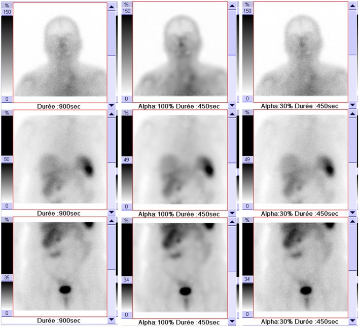
All patients with neuroendocrine tumors (NETs) disease who were referred for SRS to our department during 1-year period from January to December 2015 were consecutively included. Three nuclear physicians independently reviewed all the data sets of images which included conventional images (CI; 15 min/view) and processed images (PI) obtained by reconstructing the first 450 s extracted data using Oncoflash software package. Image analysis using a 3-point rating scale for abnormal uptake of 111 Indium-DTPA-Phe-octreotide in any lesion or organ was interpreted as positive, uncertain, or negative for the evidence of NET disease. A maximum grade uptake of the radiotracer in the lesion was assessed by the Krenning scale method. The results of image interpretation by the two methods were considered significantly discordant when the difference in organ involvement assessment was negative vs. positive or in lesion uptake was ≥2 grades. Agreement between the results of two methods and by different scan observers was evaluated using Cohen κ coefficients.
There was no significant ( = 0.403) correlation between data acquisition protocol and quality image. The rates of significant discrepancies for exam interpretation and organs involvement assessment were 2.8 and 2.6%, respectively. Mean κ values revealed a good agreement for concordance between CI and PI interpretation without difference of agreement for inter/intra-observer analysis.
Our results suggest the feasibility to use a Pixon-based reconstruction method for SRS planar images allowing a twofold reduction of acquisition time and without significant alteration of image quality or on image interpretation.
本研究的目的是评估基于像素的重建方法对平面生长抑素受体闪烁显像(SRS)的诊断效能。
连续纳入2015年1月至12月期间因SRS转诊至我科的所有神经内分泌肿瘤(NETs)患者。三名核医学医师独立审查所有图像数据集,其中包括常规图像(CI;15分钟/视图)和通过使用Oncoflash软件包重建提取的前450秒数据获得的处理后图像(PI)。使用3分制评分量表对任何病变或器官中111铟-二乙三胺五乙酸-苯丙氨酸-奥曲肽的异常摄取进行图像分析,结果解释为NET疾病证据呈阳性、不确定或阴性。通过Krenning量表法评估病变中放射性示踪剂的最大摄取等级。当器官受累评估的差异为阴性与阳性或病变摄取差异≥2级时,两种方法的图像解释结果被认为存在显著不一致。使用Cohen κ系数评估两种方法以及不同扫描观察者结果之间的一致性。
数据采集方案与图像质量之间无显著(=0.403)相关性。检查解释和器官受累评估的显著差异率分别为2.8%和2.6%。平均κ值显示CI和PI解释之间的一致性良好,观察者间/观察者内分析的一致性无差异。
我们的结果表明,基于像素的重建方法用于SRS平面图像是可行的,可使采集时间减少一半,且图像质量或图像解释无显著改变。