Lujan Pablo, Rubio Teresa, Varsano Giulia, Köhn Maja
Genome Biology Unit, European Molecular Biology Laboratory, Heidelberg, Germany.
Centre for Biological Signalling Studies (BIOSS), University of Freiburg, Freiburg, Germany.
Commun Integr Biol. 2017 Jul 5;10(4):e1338990. doi: 10.1080/19420889.2017.1338990. eCollection 2017.
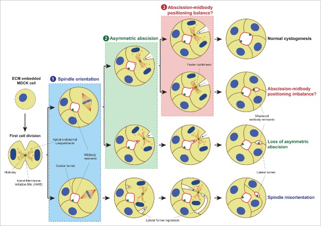
The maintenance of the epithelial architecture during tissue proliferation is achieved by apical positioning of the midbody after cell division. Consequently, midbody mislocalization contributes to epithelial architecture disruption, a fundamental event during epithelial tumorigenesis. Studies in 3D polarized epithelial MDCK or Caco2 cell models, where midbody misplacement leads to multiple ectopic but fully polarized lumen-containing cysts, revealed that this phenotype can be caused by 2 different scenarios: the loss of mitotic spindle orientation or the loss of asymmetric abscission. In addition, we have recently proposed a third cellular mechanism where the midbody mislocalization is achieved through cytokinesis acceleration driven by the cancer-promoting phosphatase of regenerating liver (PRL)-3. Here we critically review these findings, and we furthermore present new data indicating that midbodies themselves might act as signal unit for polarization since they can infer apical characteristics to a basal membrane.
在组织增殖过程中,上皮结构的维持是通过细胞分裂后中间体的顶端定位来实现的。因此,中间体定位错误会导致上皮结构破坏,这是上皮肿瘤发生过程中的一个基本事件。在三维极化上皮MDCK或Caco2细胞模型中的研究表明,中间体错位会导致多个异位但完全极化的含管腔囊肿,该表型可能由两种不同情况引起:有丝分裂纺锤体方向的丧失或不对称分裂的丧失。此外,我们最近提出了第三种细胞机制,即通过促进癌症的再生肝脏磷酸酶(PRL)-3驱动的胞质分裂加速来实现中间体定位错误。在此,我们批判性地回顾了这些发现,并且进一步展示了新的数据,表明中间体本身可能作为极化的信号单元,因为它们可以将顶端特征传递给基底膜。