• 文献检索
  • 文档翻译
  • 深度研究
  • 学术资讯
  • Suppr Zotero 插件Zotero 插件
  • 邀请有礼
  • 套餐&价格
  • 历史记录
应用&插件
Suppr Zotero 插件Zotero 插件浏览器插件Mac 客户端Windows 客户端微信小程序
定价
高级版会员购买积分包购买API积分包
服务
文献检索文档翻译深度研究API 文档MCP 服务
关于我们
关于 Suppr公司介绍联系我们用户协议隐私条款
关注我们

Suppr 超能文献

核心技术专利:CN118964589B侵权必究
粤ICP备2023148730 号-1Suppr @ 2026

文献检索

告别复杂PubMed语法,用中文像聊天一样搜索,搜遍4000万医学文献。AI智能推荐,让科研检索更轻松。

立即免费搜索

文件翻译

保留排版,准确专业,支持PDF/Word/PPT等文件格式,支持 12+语言互译。

免费翻译文档

深度研究

AI帮你快速写综述,25分钟生成高质量综述,智能提取关键信息,辅助科研写作。

立即免费体验

数字化牙颌模型与锥形束 CT 扫描的整合:一项临床验证研究。

Integration of digital dental casts in cone beam computed tomography scans-a clinical validation study.

机构信息

Department of Orthodontics and Craniofacial Biology, Radboud University Medical Centre, 309 Dentistry, PO Box 9101, 6500 HB, Nijmegen, The Netherlands.

Department of Oral and Maxillofacial Surgery, Radboud University Medical Centre, Nijmegen, The Netherlands.

出版信息

Clin Oral Investig. 2018 Apr;22(3):1215-1222. doi: 10.1007/s00784-017-2203-2. Epub 2017 Sep 20.

DOI:10.1007/s00784-017-2203-2
PMID:28932947
原文链接:https://pmc.ncbi.nlm.nih.gov/articles/PMC5866842/
Abstract

OBJECTIVES

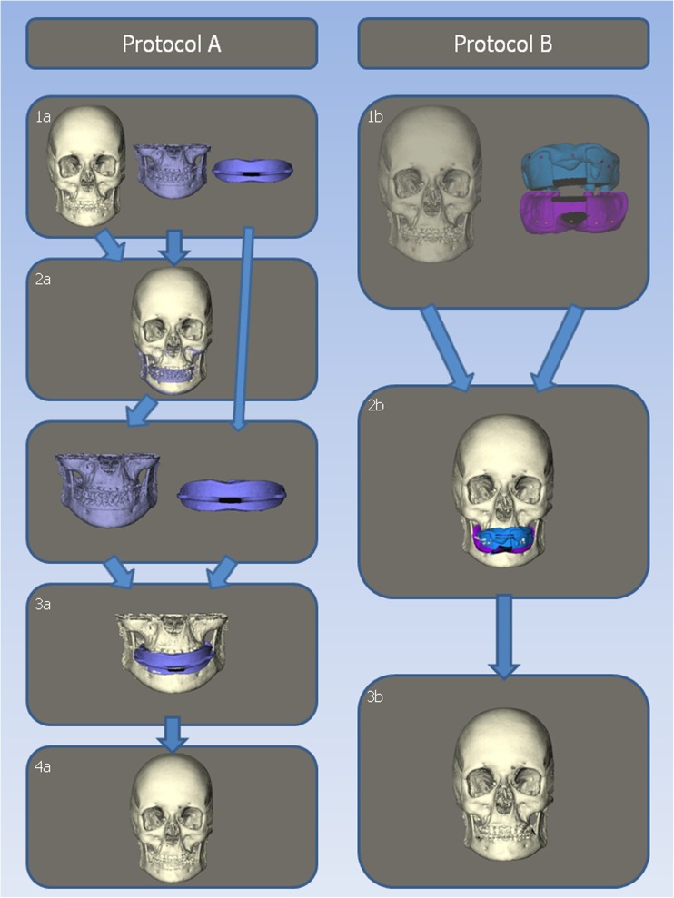
Images derived from cone beam computed tomography (CBCT) scans lack detailed information on the dentition and interocclusal relationships needed for proper surgical planning and production of surgical splints. To get a proper representation of the dentition, integration of a digital dental model into the CBCT scan is necessary. The aim of this study was to validate a simplified protocol to integrate digital dental models into CBCT scans using only one scan.

MATERIALS AND METHODS

Conventional protocol A used one combined upper and lower impression and two CBCT scans. The new protocol B included placement of ten markers on the gingiva, one CBCT scan, and two separate impressions of the upper and lower dentition. Twenty consecutive patients, scheduled for mandibular advancement surgery, were included. To validate protocol B, 3-dimensional reconstructions were made, which were compared by calculating the mean intersurface distances obtained with both protocols.

RESULTS

The mean distance for all patients for the upper jaw is 0.39 mm and for the lower jaw is 0.30 mm. For ten out of 20 patients, all distances were less than 1 mm. For the other ten patients, all distances were less than 2 mm.

CONCLUSIONS

Mean distances of 0.39 and 0.30 mm are clinically acceptable and comparable to other studies; therefore, this new protocol is clinically accurate.

CLINICAL RELEVANCE

This new protocol seems to be clinically accurate. It is less time consuming, gives less radiation exposure for the patient, and has a lower risk for positional errors of the impressions compared to other integration protocols.

摘要

目的

来自锥形束 CT(CBCT)扫描的图像缺乏详细的牙列和咬合关系信息,这对于正确的手术规划和手术夹板的制作是必要的。为了获得牙列的适当表示,需要将数字牙科模型集成到 CBCT 扫描中。本研究的目的是验证一种使用单次扫描将数字牙科模型集成到 CBCT 扫描中的简化方案。

材料和方法

常规方案 A 使用一个上下颌联合印模和两个 CBCT 扫描。新方案 B 包括在上牙龈上放置十个标记,一个 CBCT 扫描,以及上下牙列的两个单独印模。连续纳入 20 名计划接受下颌前伸手术的患者。为了验证方案 B,制作了三维重建,并通过计算两种方案获得的平均表面距离来比较。

结果

所有患者的上颌平均距离为 0.39mm,下颌为 0.30mm。20 名患者中有 10 名患者的所有距离均小于 1mm,另外 10 名患者的所有距离均小于 2mm。

结论

平均距离为 0.39 和 0.30mm 在临床上是可接受的,与其他研究相当;因此,这种新方案在临床上是准确的。

临床意义

这种新方案似乎在临床上是准确的。与其他整合方案相比,它的耗时更少,对患者的辐射暴露更少,并且印模的位置误差风险更低。

https://cdn.ncbi.nlm.nih.gov/pmc/blobs/4441/5866842/b609ed656df5/784_2017_2203_Fig3_HTML.jpg
https://cdn.ncbi.nlm.nih.gov/pmc/blobs/4441/5866842/c325d6b33ce7/784_2017_2203_Fig2_HTML.jpg
https://cdn.ncbi.nlm.nih.gov/pmc/blobs/4441/5866842/b609ed656df5/784_2017_2203_Fig3_HTML.jpg
https://cdn.ncbi.nlm.nih.gov/pmc/blobs/4441/5866842/c325d6b33ce7/784_2017_2203_Fig2_HTML.jpg
https://cdn.ncbi.nlm.nih.gov/pmc/blobs/4441/5866842/b609ed656df5/784_2017_2203_Fig3_HTML.jpg

相似文献

1
Integration of digital dental casts in cone beam computed tomography scans-a clinical validation study.数字化牙颌模型与锥形束 CT 扫描的整合:一项临床验证研究。
Clin Oral Investig. 2018 Apr;22(3):1215-1222. doi: 10.1007/s00784-017-2203-2. Epub 2017 Sep 20.
2
A novel method for fusion of intra-oral scans and cone-beam computed tomography scans for orthognathic surgery planning.一种用于正颌外科手术规划的口腔内扫描与锥形束计算机断层扫描融合的新方法。
J Craniomaxillofac Surg. 2016 Feb;44(2):160-6. doi: 10.1016/j.jcms.2015.11.017. Epub 2015 Dec 8.
3
A cone-beam computed tomography triple scan procedure to obtain a three-dimensional augmented virtual skull model appropriate for orthognathic surgery planning.一种用于获取适用于正颌外科手术规划的三维增强虚拟颅骨模型的锥形束计算机断层扫描三重扫描程序。
J Craniofac Surg. 2009 Mar;20(2):297-307. doi: 10.1097/SCS.0b013e3181996803.
4
Validation of a new method for building a three-dimensional physical model of the skull and dentition.一种构建颅骨和牙列三维物理模型新方法的验证
Br J Oral Maxillofac Surg. 2012 Jan;50(1):49-54. doi: 10.1016/j.bjoms.2010.11.009. Epub 2010 Dec 30.
5
Virtual surgical planning for orthognathic surgery using digital data transfer and an intraoral fiducial marker: the charlotte method.使用数字数据传输和口内基准标记进行正颌外科手术的虚拟手术规划:夏洛特方法
J Oral Maxillofac Surg. 2015 Jun;73(6):1143-58. doi: 10.1016/j.joms.2014.12.008. Epub 2014 Dec 13.
6
Replacement of the Distorted Dentition of the Cone-Beam Computed Tomography Scans for Orthognathic Surgery Planning.用于正颌外科手术规划的锥形束计算机断层扫描中变形牙列的替换
J Oral Maxillofac Surg. 2018 Jul;76(7):1561.e1-1561.e8. doi: 10.1016/j.joms.2018.02.018. Epub 2018 Feb 28.
7
Integration of digital dental casts in cone-beam computed tomography scans.数字牙模在锥形束计算机断层扫描中的整合。
ISRN Dent. 2012;2012:949086. doi: 10.5402/2012/949086. Epub 2012 Sep 23.
8
A cone-beam CT based technique to augment the 3D virtual skull model with a detailed dental surface.一种基于锥束CT的技术,用于用详细的牙齿表面增强三维虚拟颅骨模型。
Int J Oral Maxillofac Surg. 2009 Jan;38(1):48-57. doi: 10.1016/j.ijom.2008.11.006. Epub 2008 Dec 31.
9
Registration accuracy in the integration of laser-scanned dental images into maxillofacial cone-beam computed tomography images.激光扫描牙颌图像与颌面锥形束 CT 图像融合中的配准精度。
Am J Orthod Dentofacial Orthop. 2011 Oct;140(4):585-91. doi: 10.1016/j.ajodo.2011.04.018.
10
Dimensional accuracy of digital dental models from cone-beam computed tomography scans of alginate impressions according to time elapsed after the impressions.根据藻酸盐印模采集后经过的时间,分析锥形束计算机断层扫描藻酸盐印模所获数字牙科模型的尺寸精度。
Am J Orthod Dentofacial Orthop. 2016 Feb;149(2):287-94. doi: 10.1016/j.ajodo.2015.08.014.

引用本文的文献

1
Accuracy and reliability of a three-dimensional superimposition method for maxillary jaws and dentition.一种用于上颌骨和牙列的三维叠加方法的准确性和可靠性。
BMC Oral Health. 2025 Jul 30;25(1):1281. doi: 10.1186/s12903-025-06637-4.
2
Three-dimensional finite element analysis of teeth displacement patterns under four anchorage designs for maxillary molar distalization using clear aligners: a real-case based simulation study.使用透明矫治器对上颌磨牙远移的四种支抗设计下牙齿位移模式的三维有限元分析:一项基于实际病例的模拟研究
BMC Oral Health. 2025 Jul 2;25(1):1019. doi: 10.1186/s12903-025-06375-7.
3
Two experimental methods to integrate intra-oral scans into 3D stereophotogrammetric facial images.

本文引用的文献

1
Diagnostic accuracy and measurement sensitivity of digital models for orthodontic purposes: A systematic review.正畸用途数字模型的诊断准确性和测量敏感性:一项系统评价。
Am J Orthod Dentofacial Orthop. 2016 Feb;149(2):161-70. doi: 10.1016/j.ajodo.2015.06.029.
2
Automatic Superimposition of Palatal Fiducial Markers for Accurate Integration of Digital Dental Model and Cone Beam Computed Tomography.用于数字牙科模型与锥形束计算机断层扫描精确整合的腭部基准标记自动叠加
J Oral Maxillofac Surg. 2015 Aug;73(8):1616.e1-10. doi: 10.1016/j.joms.2015.04.004. Epub 2015 Apr 14.
3
Virtual surgical planning for orthognathic surgery using digital data transfer and an intraoral fiducial marker: the charlotte method.
将口腔内扫描整合到三维立体摄影面部图像中的两种实验方法。
Clin Oral Investig. 2025 Jan 9;29(1):54. doi: 10.1007/s00784-024-06138-8.
4
Accuracy evaluation of dental CBCT and scanned model registration method based on pulp horn mapping surface: an in vitro proof-of-concept.基于牙髓角映射面的口腔锥形束 CT 与扫描模型配准方法的准确性评估:体外概念验证。
BMC Oral Health. 2024 Jul 22;24(1):827. doi: 10.1186/s12903-024-04565-3.
5
Digital registration versus cone-beam computed tomography for evaluating implant position: a prospective cohort study.数字化印模与锥形束 CT 用于评估种植体位置的比较:一项前瞻性队列研究。
BMC Oral Health. 2024 Mar 4;24(1):304. doi: 10.1186/s12903-024-04088-x.
6
[Research progress of digital occlusion setup in orthognathic surgery].[正颌外科数字化咬合设计的研究进展]
Zhongguo Xiu Fu Chong Jian Wai Ke Za Zhi. 2023 Feb 15;37(2):247-251. doi: 10.7507/1002-1892.202210086.
7
Evaluation of the impact of reference tooth morphology and alignment on model measurement accuracy.评估参考牙齿形态和排列对模型测量准确性的影响。
Ann Transl Med. 2022 Jun;10(12):670. doi: 10.21037/atm-22-2497.
8
Accuracy of a surface-based fusion method when integrating digital models and the cone beam computed tomography scans with metal artifacts.基于表面的融合方法在整合数字模型和带有金属伪影的锥形束 CT 扫描时的准确性。
Sci Rep. 2022 May 16;12(1):8034. doi: 10.1038/s41598-022-11677-9.
9
Three-dimensional inlay-guided endodontics applied in variant root canals: A case report and review of literature.三维嵌体引导下牙髓治疗应用于变异根管:1例病例报告及文献复习
World J Clin Cases. 2021 Dec 26;9(36):11425-11436. doi: 10.12998/wjcc.v9.i36.11425.
10
Integration of imaging modalities in digital dental workflows - possibilities, limitations, and potential future developments.数字化牙科工作流程中影像学整合的可能性、局限性及潜在未来发展。
Dentomaxillofac Radiol. 2021 Oct 1;50(7):20210268. doi: 10.1259/dmfr.20210268. Epub 2021 Sep 14.
使用数字数据传输和口内基准标记进行正颌外科手术的虚拟手术规划:夏洛特方法
J Oral Maxillofac Surg. 2015 Jun;73(6):1143-58. doi: 10.1016/j.joms.2014.12.008. Epub 2014 Dec 13.
4
Intra-arch dimensional measurement validity of laser-scanned digital dental models compared with the original plaster models: a systematic review.激光扫描数字牙模与原始石膏牙模的牙弓内尺寸测量有效性:一项系统评价
Orthod Craniofac Res. 2015 May;18(2):65-76. doi: 10.1111/ocr.12068. Epub 2015 Feb 11.
5
Generation of virtual models for planning orthognathic surgery using a modified multimodal image fusion technique.使用改良的多模态图像融合技术生成用于正颌外科手术规划的虚拟模型。
Int J Oral Maxillofac Surg. 2015 Apr;44(4):462-9. doi: 10.1016/j.ijom.2014.11.007. Epub 2014 Dec 2.
6
Reproducibility and accuracy of linear measurements on dental models derived from cone-beam computed tomography compared with digital dental casts.锥形束计算机断层扫描获取的牙模与数字牙颌模型的线性测量的可重复性和准确性比较。
Am J Orthod Dentofacial Orthop. 2014 Sep;146(3):328-36. doi: 10.1016/j.ajodo.2014.05.026.
7
Integration accuracy of laser-scanned dental models into maxillofacial cone beam computed tomography images of different voxel sizes with different segmentation threshold settings.在不同体素大小及不同分割阈值设置下,激光扫描牙科模型与颌面锥形束计算机断层扫描图像的整合精度。
Oral Surg Oral Med Oral Pathol Oral Radiol. 2014 Jun;117(6):780-6. doi: 10.1016/j.oooo.2014.02.022. Epub 2014 Mar 12.
8
A novel approach for planning orthognathic surgery: the integration of dental casts into three-dimensional printed mandibular models.一种正颌外科手术规划的新方法:将牙模整合到三维打印的下颌模型中。
Int J Oral Maxillofac Surg. 2014 Apr;43(4):454-9. doi: 10.1016/j.ijom.2013.08.016. Epub 2013 Oct 3.
9
Digital replacement of the distorted dentition acquired by cone beam computed tomography (CBCT): a pilot study.基于锥形束 CT(CBCT)获取的失真牙列的数字置换:一项初步研究。
Int J Oral Maxillofac Surg. 2013 Nov;42(11):1488-93. doi: 10.1016/j.ijom.2013.01.026. Epub 2013 May 2.
10
Integration of digital dental casts in cone-beam computed tomography scans.数字牙模在锥形束计算机断层扫描中的整合。
ISRN Dent. 2012;2012:949086. doi: 10.5402/2012/949086. Epub 2012 Sep 23.