Department of Ultrasound, The Second Affiliated Hospital of Soochow University, Suzhou, Jiangsu 215004, China.
Department of Psychiatry, The Second Affiliated Hospital of Soochow University, Suzhou, Jiangsu 215004, China.
Chin Med J (Engl). 2017 Oct 5;130(19):2291-2295. doi: 10.4103/0366-6999.215329.
Numerous studies have demonstrated that patients with Parkinson's disease (PD) have a higher prevalence of substantia nigra (SN) hyperechogenicity compared with controls. Our aim was to explore the neuroimaging characteristics of transcranial sonography (TCS) of patients with PD and those with PD with dementia (PDD). The correlation between the echogenicity of the SN and clinical symptoms in Chinese patients with PDD was also assessed.
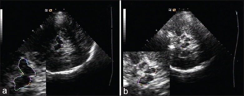
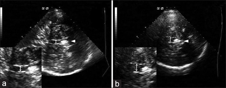
The ratios of SN hyperechogenicity (SN+), maximum sizes of SN+, and widths of third ventricle (TV) were measured using TCS for all the recruited patients. Data were analyzed using one-way analysis of variance, rank-sum test, Chi-square test, and receiver-operating characteristic (ROC) curve analysis.
The final statistical analysis included 46 PDD patients, 52 PD patients, and 40 controls. There were no significant differences in ratios of SN+ and maximum sizes of SN+ between PDD and PD groups (P > 0.05). TV widths were significantly larger in PDD group (7.1 ± 1.9 mm) than in PD group (6.0 ± 2.0 mm) and controls (5.9 ± 1.5 mm, P < 0.05); however, the ratios of enlarged TV did not differ among the three groups (P = 0.059). When cutoff value was set at 6.8 mm, the TV width had a relatively high sensitivity and specificity in discriminating between PDD and PD groups (P = 0.030) and between PDD group and controls (P = 0.003), based on ROC curve analysis. In PDD patients, SN+ was more frequently detected in akinetic-rigid subgroup, and patients with SN+ showed significantly higher Hoehn and Yahr stage and Nonmotor Symptoms Questionnaire scores (P < 0.05).
Compared to Chinese patients with PD, patients with PDD had a wider TV, altered SN sonographic features, and more severe clinical symptoms. Our findings suggest that TCS can be used to assess brain atrophy in PD and may be useful in discriminating between PD with and without dementia.
大量研究表明,帕金森病(PD)患者的黑质(SN)回声增强发生率高于对照组。我们的目的是探讨中国 PD 患者和 PD 合并痴呆(PDD)患者经颅超声(TCS)的神经影像学特征。还评估了 PDD 患者 SN 回声强度与临床症状之间的相关性。
使用 TCS 测量所有入组患者的 SN 回声增强比(SN+)、SN+最大尺寸和第三脑室(TV)宽度。采用单因素方差分析、秩和检验、卡方检验和受试者工作特征(ROC)曲线分析进行数据分析。
最终的统计分析纳入了 46 例 PDD 患者、52 例 PD 患者和 40 例对照组。PDD 组与 PD 组 SN+比和 SN+最大尺寸无显著差异(P>0.05)。PDD 组 TV 宽度明显大于 PD 组(7.1±1.9mm)和对照组(5.9±1.5mm,P<0.05),但三组间扩大的 TV 比无差异(P=0.059)。当截断值设为 6.8mm 时,根据 ROC 曲线分析,TV 宽度在区分 PDD 与 PD 组(P=0.030)和 PDD 与对照组(P=0.003)方面具有较高的敏感性和特异性。在 PDD 患者中,运动不能僵硬亚组更常检测到 SN+,且 SN+患者的 Hoehn 和 Yahr 分期和非运动症状问卷评分显著更高(P<0.05)。
与中国 PD 患者相比,PDD 患者的 TV 更宽,SN 超声特征改变,且临床症状更严重。我们的研究结果表明,TCS 可用于评估 PD 患者的脑萎缩,有助于区分 PD 伴或不伴痴呆。