Movement Disorders Unit, Neurology Service, Complex Hospitalari Moisès Broggi, Barcelona, Spain.
Movement Disorders Unit, Neurology Service. Hospital Universitari Germans Trias i Pujol, Badalona, Barcelona, Spain.
Alzheimers Res Ther. 2024 Oct 15;16(1):227. doi: 10.1186/s13195-024-01590-w.
Diagnosis of dementia with Lewy bodies (DLB) is challenging, especially in the earlier stages of the disease, owing to the clinical overlap with other neurodegenerative diseases such as Alzheimer's (AD) and Parkinson's disease (PD). We aimed to identify the transcranial sonography (TCS) parameters that can help us to detect early DLB patients.
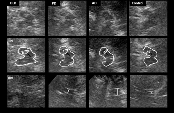
In this cross-sectional study, we prospectively recruited newly diagnosed DLB patients with less than 3 years from the onset of cognitive symptoms. For comparison purposes, we also included AD and PD patients, with a disease duration of less than 3 years, and a control group. TCS was performed to assess the substantia nigra (SN) echogenicity, the width of the third ventricle, and the frontal horns of the lateral ventricles. Subsequently, TCS images were analyzed with the medical image viewer Horos in order to quantify the intensity of the echogenicity of the SN. Univariate analysis and a logistic regression model were used to identify which variables can predict the diagnosis of DLB.
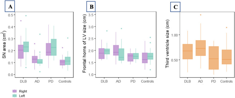
One hundred and seven participants were included (23 DLB, 26 AD, 27 PD and 31 controls). The median age of DLB patients was 75(72-77) years, with a disease duration of 2 years. DLB and PD patients showed higher SN hyperechogenicity rates (72.73% and 81.82%, respectively) and a greater area of the SN compared to AD patients and controls (p < 0.001). DLB and AD patients had wider ventricular systems than the other study groups. The SN hyperechogenicity predicted a diagnosis of DLB with an odds ratio of 22.67 (95%CI 3.98; 129.12, p < 0.001) when compared to AD patients. Unilateral and bilateral widened frontal horns predicted diagnosis of DLB compared to PD with an odds ratio of 9.5 (95%CI 0.97; 92.83, p = 0.053) and 5.7 (95%CI 0.97; 33.6, p = 0.054), respectively.
Echogenicity of the SN and widening of the frontal horns of lateral ventricles can predict the diagnosis of early DLB in this cohort of newly diagnosed patients, when compared to AD and PD patients. Transcranial sonography, a non-invasive tool, could be helpful for the diagnosis of DLB at its earlier stages.
由于与其他神经退行性疾病(如阿尔茨海默病(AD)和帕金森病(PD))的临床重叠,路易体痴呆(DLB)的诊断具有挑战性,尤其是在疾病的早期阶段。我们旨在确定有助于我们发现早期 DLB 患者的经颅超声(TCS)参数。
在这项前瞻性的横断面研究中,我们招募了从认知症状出现不到 3 年的新诊断为 DLB 的患者。为了比较目的,我们还纳入了疾病持续时间少于 3 年的 AD 和 PD 患者,以及对照组。对患者进行 TCS 检查以评估黑质(SN)回声强度、第三脑室宽度和侧脑室额角。随后,使用医学图像查看器 Horos 对 TCS 图像进行分析,以定量评估 SN 回声强度。采用单变量分析和逻辑回归模型来识别哪些变量可以预测 DLB 的诊断。
共纳入 107 名参与者(23 名 DLB、26 名 AD、27 名 PD 和 31 名对照组)。DLB 患者的中位年龄为 75(72-77)岁,疾病持续时间为 2 年。与 AD 患者和对照组相比,DLB 和 PD 患者的 SN 高回声率更高(分别为 72.73%和 81.82%),SN 面积更大(p<0.001)。DLB 和 AD 患者的脑室系统比其他研究组更宽。与 AD 患者相比,SN 高回声预测 DLB 诊断的优势比为 22.67(95%CI 3.98;129.12,p<0.001)。与 PD 患者相比,单侧和双侧额角增宽预测 DLB 诊断的优势比分别为 9.5(95%CI 0.97;92.83,p=0.053)和 5.7(95%CI 0.97;33.6,p=0.054)。
与 AD 和 PD 患者相比,在本队列中,新诊断的早期 DLB 患者 SN 的回声强度和侧脑室额角增宽可预测 DLB 的诊断。作为一种非侵入性工具,经颅超声可能有助于在疾病的早期阶段诊断 DLB。