• 文献检索
  • 文档翻译
  • 深度研究
  • 学术资讯
  • Suppr Zotero 插件Zotero 插件
  • 邀请有礼
  • 套餐&价格
  • 历史记录
应用&插件
Suppr Zotero 插件Zotero 插件浏览器插件Mac 客户端Windows 客户端微信小程序
定价
高级版会员购买积分包购买API积分包
服务
文献检索文档翻译深度研究API 文档MCP 服务
关于我们
关于 Suppr公司介绍联系我们用户协议隐私条款
关注我们

Suppr 超能文献

核心技术专利:CN118964589B侵权必究
粤ICP备2023148730 号-1Suppr @ 2026

文献检索

告别复杂PubMed语法,用中文像聊天一样搜索,搜遍4000万医学文献。AI智能推荐,让科研检索更轻松。

立即免费搜索

文件翻译

保留排版,准确专业,支持PDF/Word/PPT等文件格式,支持 12+语言互译。

免费翻译文档

深度研究

AI帮你快速写综述,25分钟生成高质量综述,智能提取关键信息,辅助科研写作。

立即免费体验

不同类型原发性局灶性肌张力障碍中基底节的超声改变:一项横断面研究。

Sonographic Alteration of Basal Ganglia in Different Forms of Primary Focal Dystonia: A Cross-sectional Study.

作者信息

Zhang Ying, Zhang Ying-Chun, Sheng Yu-Jing, Chen Xiao-Fang, Wang Cai-Shan, Ma Qi, Chen Han-Bing, Yu Li-Fang, Mao Cheng-Jie, Xiong Kang-Ping, Luo Wei-Feng, Liu Chun-Feng

机构信息

Department of Ultrasound, The Second Affiliated Hospital of Soochow University, Suzhou, Jiangsu 215004, China.

出版信息

Chin Med J (Engl). 2016 Apr 20;129(8):942-5. doi: 10.4103/0366-6999.179792.

DOI:10.4103/0366-6999.179792
PMID:27064039
原文链接:https://pmc.ncbi.nlm.nih.gov/articles/PMC4831529/
Abstract

BACKGROUND

Few studies have addressed whether abnormalities in the lenticular nucleus (LN) are characteristic transcranial sonography (TCS) echo features in patients with primary dystonia. This study aimed to explore alterations in the basal ganglia in different forms of primary focal dystonia.

METHODS

cross-sectional observational study was performed between December 2013 and December 2014 in 80 patients with different forms of primary focal dystonia and 55 neurologically normal control subjects. TCS was performed in patients and control subjects. Multiple comparisons of multiple rates were used to compare LN hyperechogenicity ratios between control and patient groups.

RESULTS

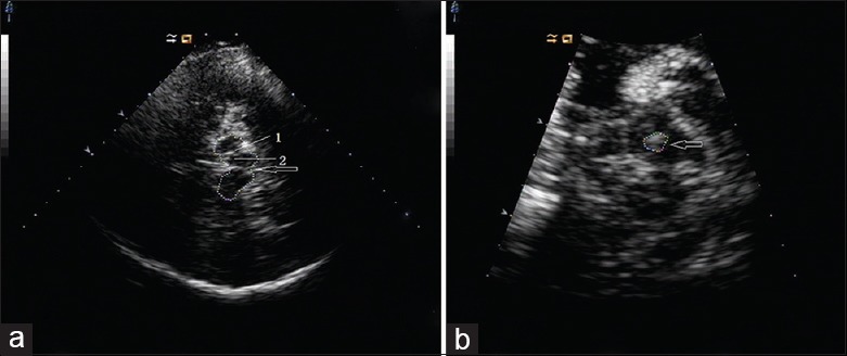
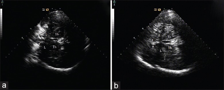
Thirteen individuals were excluded due to poor temporal bone windows, and two subjects were excluded due to disagreement in evaluation by sonologists. Totally, 70 patients (cervical dystonia, n = 30; blepharospasm, n = 30; oromandibular dystonia, n = 10) and 50 normal controls were included in the final analysis. LN hyperechogenicity was observed in 51% (36/70) of patients with primary focal dystonia, compared with 12% (6/50) of controls (P < 0.001). Substantia nigra hyperechogenicity did not differ between the two groups. LN hyperechogenicity was observed in 73% (22/30) of patients with cervical dystonia, a greater prevalence than in patients with blepharospasm (33%, 10/30, P = 0.002) and oromandibular dystonia (40%, 4/10, P = 0.126). LN hyperechogenicity was more frequently observed in patients with cervical dystonia compared with controls (73% vs. 12%, P < 0.001); however, no significant difference was detected in patients with blepharospasm (33% vs. 12%, P = 0.021) or oromandibular dystonia (40% vs. 12%, P = 0.088).

CONCLUSIONS

LN hyperechogenicity is more frequently observed in patients with primary focal dystonia than in controls. It does not appear to be a characteristic TCS echo feature in patients with blepharospasm or oromandibular dystonia.

摘要

背景

很少有研究探讨豆状核(LN)异常是否是原发性肌张力障碍患者经颅超声检查(TCS)的特征性回声表现。本研究旨在探讨不同类型原发性局灶性肌张力障碍患者基底节的改变。

方法

2013年12月至2014年12月,对80例不同类型原发性局灶性肌张力障碍患者和55例神经功能正常的对照者进行了横断面观察研究。对患者和对照者进行了TCS检查。采用多个率的多重比较来比较对照组和患者组之间的LN高回声率。

结果

13例因颞骨窗不佳被排除,2例因超声科医生评估意见不一致被排除。最终分析纳入70例患者(颈部肌张力障碍30例、眼睑痉挛30例、口下颌肌张力障碍10例)和50例正常对照者。原发性局灶性肌张力障碍患者中51%(36/70)观察到LN高回声,而对照组为12%(6/50)(P<0.001)。两组之间黑质高回声无差异。颈部肌张力障碍患者中73%(22/30)观察到LN高回声,其患病率高于眼睑痉挛患者(33%,10/30,P = 0.002)和口下颌肌张力障碍患者(40%,4/10,P = 0.126)。与对照组相比,颈部肌张力障碍患者更常观察到LN高回声(73%比12%,P<0.001);然而,眼睑痉挛患者(33%比12%,P = 0.021)或口下颌肌张力障碍患者(40%比12%,P = 0.088)未检测到显著差异。

结论

原发性局灶性肌张力障碍患者比对照组更常观察到LN高回声。它似乎不是眼睑痉挛或口下颌肌张力障碍患者的特征性TCS回声表现。

https://cdn.ncbi.nlm.nih.gov/pmc/blobs/4246/4831529/d5928ab34b13/CMJ-129-942-g002.jpg
https://cdn.ncbi.nlm.nih.gov/pmc/blobs/4246/4831529/2497ae058a3f/CMJ-129-942-g001.jpg
https://cdn.ncbi.nlm.nih.gov/pmc/blobs/4246/4831529/d5928ab34b13/CMJ-129-942-g002.jpg
https://cdn.ncbi.nlm.nih.gov/pmc/blobs/4246/4831529/2497ae058a3f/CMJ-129-942-g001.jpg
https://cdn.ncbi.nlm.nih.gov/pmc/blobs/4246/4831529/d5928ab34b13/CMJ-129-942-g002.jpg

相似文献

1
Sonographic Alteration of Basal Ganglia in Different Forms of Primary Focal Dystonia: A Cross-sectional Study.不同类型原发性局灶性肌张力障碍中基底节的超声改变:一项横断面研究。
Chin Med J (Engl). 2016 Apr 20;129(8):942-5. doi: 10.4103/0366-6999.179792.
2
Basal ganglia hyperechogenicity does not distinguish between patients with primary dystonia and healthy individuals.基底节区回声增强并不能区分原发性肌张力障碍患者和健康个体。
J Neurol. 2011 Apr;258(4):590-5. doi: 10.1007/s00415-010-5795-x. Epub 2010 Nov 2.
3
Sonographic alteration of lenticular nucleus in focal task-specific dystonia of musicians.音乐家局限性任务相关肌张力障碍的脑纹状体超声改变。
Neurodegener Dis. 2012;9(2):99-103. doi: 10.1159/000330712. Epub 2011 Sep 10.
4
Lenticular nucleus lesion in idiopathic dystonia detected by transcranial sonography.经颅超声检查发现特发性肌张力障碍中的豆状核病变。
Neurology. 1996 Nov;47(5):1284-90. doi: 10.1212/wnl.47.5.1284.
5
Sonographic detection of basal ganglia abnormalities in spasmodic dysphonia.痉挛性发声障碍中基底节异常的超声检测
Eur J Neurol. 2014 Feb;21(2):349-52. doi: 10.1111/ene.12151. Epub 2013 Apr 14.
6
Transcranial sonography in dystonia.经颅超声在肌张力障碍中的应用。
Int Rev Neurobiol. 2010;90:179-87. doi: 10.1016/S0074-7742(10)90013-5.
7
Lentiform Nucleus Hyperechogenicity in Parkinsonian Syndromes: A Systematic Review and Meta-Analysis with Consideration of Molecular Pathology.苍白球高回声在帕金森综合征中的表现:一项系统性综述和荟萃分析,同时考虑了分子病理学。
Cells. 2019 Dec 18;9(1):2. doi: 10.3390/cells9010002.
8
Sonographic detection of basal ganglia lesions in asymptomatic and symptomatic Wilson disease.超声检查对无症状和有症状肝豆状核变性患者基底节病变的检测
Neurology. 2005 May 24;64(10):1726-32. doi: 10.1212/01.WNL.0000161847.46465.B9.
9
Sonographic alteration of substantia nigra is related to parkinsonism-predominant course of X-linked dystonia-parkinsonism.黑质的超声改变与X连锁肌张力障碍-帕金森综合征以帕金森症状为主的病程相关。
Parkinsonism Relat Disord. 2017 Apr;37:43-49. doi: 10.1016/j.parkreldis.2017.01.006. Epub 2017 Jan 10.
10
[The neuroimaging features of Parkinson's disease and dystonia by transcranial sonography].[经颅超声检查帕金森病和肌张力障碍的神经影像学特征]
Zhonghua Yi Xue Za Zhi. 2015 Apr 21;95(15):1135-9.

引用本文的文献

1
Diabetic Striatopathy: Parenchymal Transcranial Sonography as a Supplement to Diagnosis at the Emergency Department.糖尿病性纹状体病:实质内经颅超声检查作为急诊科诊断的补充手段
Diagnostics (Basel). 2022 Nov 17;12(11):2838. doi: 10.3390/diagnostics12112838.
2
Is Generalized and Segmental Dystonia Accompanied by Impairments in the Dopaminergic System?全身性和节段性肌张力障碍是否伴有多巴胺能系统损害?
Front Neurol. 2021 Nov 18;12:751434. doi: 10.3389/fneur.2021.751434. eCollection 2021.
3
Echogenicity Changes in Brainstem Raphe Detected by Transcranial Parenchymal Sonography and Clinical Characteristics in Parkinson's Disease.

本文引用的文献

1
Transcranial sonography (TCS) of brain parenchyma in movement disorders: quality standards, diagnostic applications and novel technologies.运动障碍中脑实质的经颅超声检查(TCS):质量标准、诊断应用及新技术
Ultraschall Med. 2014 Aug;35(4):322-31. doi: 10.1055/s-0033-1356415. Epub 2014 Apr 24.
2
The anatomical basis of dystonia: current view using neuroimaging.运用神经影像学研究扭转痉挛的解剖学基础:当前观点
Mov Disord. 2013 Jun 15;28(7):944-57. doi: 10.1002/mds.25527.
3
Sonographic detection of basal ganglia abnormalities in spasmodic dysphonia.
经颅实质超声检测帕金森病中脑桥中缝核回声变化及临床特征
Front Neurol. 2020 Aug 7;11:821. doi: 10.3389/fneur.2020.00821. eCollection 2020.
4
Transcranial Sonographic Alterations of Substantia Nigra and Third Ventricle in Parkinson's Disease with or without Dementia.帕金森病伴或不伴痴呆患者黑质和第三脑室经颅超声改变。
Chin Med J (Engl). 2017 Oct 5;130(19):2291-2295. doi: 10.4103/0366-6999.215329.
痉挛性发声障碍中基底节异常的超声检测
Eur J Neurol. 2014 Feb;21(2):349-52. doi: 10.1111/ene.12151. Epub 2013 Apr 14.
4
Assessment of substantia nigra echogenicity in German and Filipino populations using a portable ultrasound system.使用便携式超声系统评估德国和菲律宾人群的黑质回声强度。
J Ultrasound Med. 2012 Feb;31(2):191-6. doi: 10.7863/jum.2012.31.2.191.
5
Sonographic alteration of lenticular nucleus in focal task-specific dystonia of musicians.音乐家局限性任务相关肌张力障碍的脑纹状体超声改变。
Neurodegener Dis. 2012;9(2):99-103. doi: 10.1159/000330712. Epub 2011 Sep 10.
6
Enlarged substantia nigra hyperechogenicity and risk for Parkinson disease: a 37-month 3-center study of 1847 older persons.黑质回声增强与帕金森病风险:一项针对1847名老年人的为期37个月的三中心研究
Arch Neurol. 2011 Jul;68(7):932-7. doi: 10.1001/archneurol.2011.141.
7
Structural, functional and molecular imaging of the brain in primary focal dystonia--a review.原发性局灶性肌张力障碍的脑结构、功能和分子影像学——综述。
Neuroimage. 2011 Jun 1;56(3):1011-20. doi: 10.1016/j.neuroimage.2011.02.045. Epub 2011 Feb 22.
8
Basal ganglia hyperechogenicity does not distinguish between patients with primary dystonia and healthy individuals.基底节区回声增强并不能区分原发性肌张力障碍患者和健康个体。
J Neurol. 2011 Apr;258(4):590-5. doi: 10.1007/s00415-010-5795-x. Epub 2010 Nov 2.
9
Hereditary dystonia as a neurodevelopmental circuit disorder: Evidence from neuroimaging.遗传性肌张力障碍作为一种神经发育性回路障碍:神经影像学证据。
Neurobiol Dis. 2011 May;42(2):202-9. doi: 10.1016/j.nbd.2010.10.010. Epub 2010 Oct 19.
10
EFNS guidelines on diagnosis and treatment of primary dystonias.EFNS 原发性运动障碍诊断和治疗指南。
Eur J Neurol. 2011 Jan;18(1):5-18. doi: 10.1111/j.1468-1331.2010.03042.x.