• 文献检索
  • 文档翻译
  • 深度研究
  • 学术资讯
  • Suppr Zotero 插件Zotero 插件
  • 邀请有礼
  • 套餐&价格
  • 历史记录
应用&插件
Suppr Zotero 插件Zotero 插件浏览器插件Mac 客户端Windows 客户端微信小程序
定价
高级版会员购买积分包购买API积分包
服务
文献检索文档翻译深度研究API 文档MCP 服务
关于我们
关于 Suppr公司介绍联系我们用户协议隐私条款
关注我们

Suppr 超能文献

核心技术专利:CN118964589B侵权必究
粤ICP备2023148730 号-1Suppr @ 2026

文献检索

告别复杂PubMed语法,用中文像聊天一样搜索,搜遍4000万医学文献。AI智能推荐,让科研检索更轻松。

立即免费搜索

文件翻译

保留排版,准确专业,支持PDF/Word/PPT等文件格式,支持 12+语言互译。

免费翻译文档

深度研究

AI帮你快速写综述,25分钟生成高质量综述,智能提取关键信息,辅助科研写作。

立即免费体验

实验性内脏利什曼病的新见解:杜氏利什曼原虫毒力的实时体内成像

New insights into experimental visceral leishmaniasis: Real-time in vivo imaging of Leishmania donovani virulence.

作者信息

Melo Guilherme D, Goyard Sophie, Lecoeur Hervé, Rouault Eline, Pescher Pascale, Fiette Laurence, Boissonnas Alexandre, Minoprio Paola, Lang Thierry

机构信息

Institut Pasteur, Laboratoire des Processus Infectieux à Trypanosomatidés, Département Infection et Epidémiologie, 25-28 rue du Dr Roux, Paris, France.

Institut Pasteur, Unité de Parasitologie Moléculaire et Signalisation, Département de Parasites et Insectes Vecteurs, 25-28 rue du Dr Roux, Paris, France.

出版信息

PLoS Negl Trop Dis. 2017 Sep 25;11(9):e0005924. doi: 10.1371/journal.pntd.0005924. eCollection 2017 Sep.

DOI:10.1371/journal.pntd.0005924
PMID:28945751
原文链接:https://pmc.ncbi.nlm.nih.gov/articles/PMC5629011/
Abstract

UNLABELLED

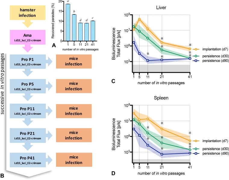
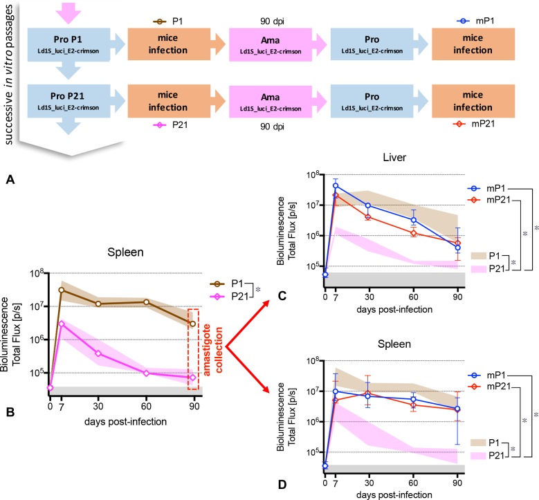
Visceral leishmaniasis is an insidious neglected disease with worldwide distribution. It is caused by parasites from the Leishmania donovani complex, which are able to be transmitted by different species of phlebotomine sand flies and to infect numerous mammal hosts. Despite the high number of people infected or at risk, and the remarkable quantity of studies focusing on this disease, a proper experimental model to efficiently decipher the infectious process of visceral leishmaniasis taking into account the nuances of parasite’s virulence and the duration of the infection is still lacking. Therefore, using golden Syrian hamsters and BALB/c mice, state-of-the-art genetic manipulation applied on a fully virulent L. donovani strain and in vivo imaging approaches, we describe herein three benefits for experimental visceral leishmaniasis: (i) the development of a double transfected bioluminescent (firefly luciferase) and fluorescent (E2-crimson) virulent strain of L. donovani (Ld1S_luci_E2-crimson), favoring a wide range of both in vivo and in vitro investigations, (ii) the establishment of a non-invasive mouse model to evaluate the infectious process during visceral leishmaniasis and the parasite’s virulence in real time, allowing longitudinal studies with the same animals, and (iii) the elaboration of a suitable method to reinstate (and verify anew) the virulence in a population of attenuated parasites, by recovering persistent parasites from chronic infected mice. Consequently, these results open up new perspectives on the study of visceral leishmaniasis, especially in the fields of therapeutics and vaccinology, since the model described herein renders now possible long-lasting follow up studies, with easy and accurate day-by-day verifications of the infection status along with a reduced number of laboratory animals.

TRIAL REGISTRATION

ClinicalTrials.gov 2013-0047.

摘要

未标注

内脏利什曼病是一种分布于全球的隐匿性被忽视疾病。它由杜氏利什曼原虫复合体的寄生虫引起,这些寄生虫可通过不同种类的白蛉传播,并感染众多哺乳动物宿主。尽管感染人数众多或面临感染风险,且针对该疾病的研究数量可观,但仍缺乏一个合适的实验模型来有效解读内脏利什曼病的感染过程,同时考虑到寄生虫毒力的细微差别和感染持续时间。因此,我们使用叙利亚金黄地鼠和BALB/c小鼠,对一种完全有毒力的杜氏利什曼原虫菌株应用最先进的基因操作技术,并采用体内成像方法,在此描述了实验性内脏利什曼病的三个优势:(i)构建了一种双转染的生物发光(萤火虫荧光素酶)和荧光(E2-深红色)的杜氏利什曼原虫有毒力菌株(Ld1S_luci_E2-深红色),有利于进行广泛的体内和体外研究;(ii)建立了一种非侵入性小鼠模型,用于实时评估内脏利什曼病的感染过程和寄生虫毒力,允许对同一动物进行纵向研究;(iii)制定了一种合适的方法,通过从慢性感染小鼠中回收持续性寄生虫,恢复(并重新验证)减毒寄生虫群体的毒力。因此,这些结果为内脏利什曼病的研究开辟了新的视角,特别是在治疗学和疫苗学领域,因为本文所述模型现在使得长期随访研究成为可能,能够轻松、准确地逐日核实感染状态,同时减少实验动物数量。

试验注册

ClinicalTrials.gov 2013 - 0047。

https://cdn.ncbi.nlm.nih.gov/pmc/blobs/57f5/5629011/754146c617c4/pntd.0005924.g006.jpg
https://cdn.ncbi.nlm.nih.gov/pmc/blobs/57f5/5629011/b69e2399f7bd/pntd.0005924.g005.jpg
https://cdn.ncbi.nlm.nih.gov/pmc/blobs/57f5/5629011/754146c617c4/pntd.0005924.g006.jpg
https://cdn.ncbi.nlm.nih.gov/pmc/blobs/57f5/5629011/b69e2399f7bd/pntd.0005924.g005.jpg
https://cdn.ncbi.nlm.nih.gov/pmc/blobs/57f5/5629011/754146c617c4/pntd.0005924.g006.jpg

相似文献

1
New insights into experimental visceral leishmaniasis: Real-time in vivo imaging of Leishmania donovani virulence.实验性内脏利什曼病的新见解:杜氏利什曼原虫毒力的实时体内成像
PLoS Negl Trop Dis. 2017 Sep 25;11(9):e0005924. doi: 10.1371/journal.pntd.0005924. eCollection 2017 Sep.
2
Imaging visceral leishmaniasis in real time with golden hamster model: Monitoring the parasite burden and hamster transcripts to further characterize the immunological responses of the host.利用金黄仓鼠模型实时成像内脏利什曼病:监测寄生虫负荷和仓鼠转录本以进一步表征宿主的免疫反应。
Parasitol Int. 2017 Feb;66(1):933-939. doi: 10.1016/j.parint.2016.10.020. Epub 2016 Oct 26.
3
[Experimental visceral leishmaniasis in golden hamsters].[金黄仓鼠实验性内脏利什曼病]
Parazitologiia. 1987 Sep-Oct;21(5):637-42.
4
In vivo characterization of two additional Leishmania donovani strains using the murine and hamster model.使用小鼠和仓鼠模型对另外两种杜氏利什曼原虫菌株进行体内特性分析。
Parasite Immunol. 2016 May;38(5):290-302. doi: 10.1111/pim.12316.
5
Evidence that a naturally occurring single nucleotide polymorphism in the RagC gene of Leishmania donovani contributes to reduced virulence.证据表明,利什曼原虫 RagC 基因中的一个自然发生的单核苷酸多态性导致其毒力降低。
PLoS Negl Trop Dis. 2021 Feb 23;15(2):e0009079. doi: 10.1371/journal.pntd.0009079. eCollection 2021 Feb.
6
Screening Leishmania donovani complex-specific genes required for visceral disease.筛查内脏利什曼病所需的杜氏利什曼原虫复合种特异性基因。
Methods Mol Biol. 2015;1201:339-61. doi: 10.1007/978-1-4939-1438-8_20.
7
Tissue Specific Dual RNA-Seq Defines Host-Parasite Interplay in Murine Visceral Leishmaniasis Caused by Leishmania donovani and Leishmania infantum.组织特异性双重 RNA-Seq 定义了由利什曼原虫和婴儿利什曼原虫引起的小鼠内脏利什曼病中的宿主-寄生虫相互作用。
Microbiol Spectr. 2022 Apr 27;10(2):e0067922. doi: 10.1128/spectrum.00679-22. Epub 2022 Apr 6.
8
Leishmaniasis in Turkey: Visceral and cutaneous leishmaniasis caused by Leishmania donovani in Turkey.土耳其的利什曼病:土耳其由杜氏利什曼原虫引起的内脏利什曼病和皮肤利什曼病。
Acta Trop. 2017 Sep;173:90-96. doi: 10.1016/j.actatropica.2017.05.032. Epub 2017 Jun 3.
9
Dermotropic Leishmania donovani in Sri Lanka: visceralizing potential in clinical and preclinical studies.斯里兰卡的嗜皮利什曼原虫杜氏利什曼原虫:临床和临床前研究中的内脏化潜力。
Parasitology. 2018 Apr;145(4):443-452. doi: 10.1017/S003118201700169X. Epub 2017 Nov 8.
10
Reduced pathogenicity of fructose-1,6-bisphosphatase deficient Leishmania donovani and its use as an attenuated strain to induce protective immunogenicity.果糖-1,6-二磷酸酶缺陷型杜氏利什曼原虫致病性降低及其作为减毒菌株诱导保护性免疫原性的研究。
Vaccine. 2018 Feb 21;36(9):1190-1202. doi: 10.1016/j.vaccine.2018.01.032. Epub 2018 Feb 1.

引用本文的文献

1
Malnutrition exacerbates pathogenesis of Lutzomyia longipalpis sand fly-transmitted Leishmania donovani.营养不良会加剧长须罗蛉传播的杜氏利什曼原虫的发病机制。
Commun Biol. 2025 May 13;8(1):746. doi: 10.1038/s42003-025-08106-8.
2
Comparison of Bioluminescent Substrates in Natural Infection Models of Neglected Parasitic Diseases.比较忽视寄生虫病自然感染模型中的生物发光底物。
Int J Mol Sci. 2022 Dec 16;23(24):16074. doi: 10.3390/ijms232416074.
3
Reporter gene systems: A powerful tool for studies.报告基因系统:研究的强大工具。

本文引用的文献

1
Unveiling Cerebral Leishmaniasis: parasites and brain inflammation in Leishmania donovani infected mice.揭示脑利什曼病:感染杜氏利什曼原虫的小鼠中的寄生虫和脑部炎症。
Sci Rep. 2017 Aug 16;7(1):8454. doi: 10.1038/s41598-017-09085-5.
2
Imaging visceral leishmaniasis in real time with golden hamster model: Monitoring the parasite burden and hamster transcripts to further characterize the immunological responses of the host.利用金黄仓鼠模型实时成像内脏利什曼病:监测寄生虫负荷和仓鼠转录本以进一步表征宿主的免疫反应。
Parasitol Int. 2017 Feb;66(1):933-939. doi: 10.1016/j.parint.2016.10.020. Epub 2016 Oct 26.
3
In vivo near-infrared fluorescence imaging of Leishmania amazonensis expressing infrared fluorescence protein (iRFP) for real-time monitoring of cutaneous leishmaniasis in mice.
Curr Res Microb Sci. 2022 Sep 29;3:100165. doi: 10.1016/j.crmicr.2022.100165. eCollection 2022.
4
The vertical transmission of Enteritidis in a One-Health context.在“同一健康”背景下肠炎沙门氏菌肠炎亚种的垂直传播
One Health. 2022 Dec 5;16:100469. doi: 10.1016/j.onehlt.2022.100469. eCollection 2023 Jun.
5
A Simple Bioluminescent Assay for the Screening of Cytotoxic Molecules Against the Intracellular Form of Leishmania infantum.一种用于筛选针对婴儿利什曼原虫细胞内形式的细胞毒性分子的简单生物发光测定法。
Methods Mol Biol. 2022;2524:127-147. doi: 10.1007/978-1-0716-2453-1_10.
6
Experimental evolution links post-transcriptional regulation to Leishmania fitness gain.实验进化将转录后调控与利什曼原虫适应性增益联系起来。
PLoS Pathog. 2022 Mar 16;18(3):e1010375. doi: 10.1371/journal.ppat.1010375. eCollection 2022 Mar.
7
Bioprospecting marine actinomycetes for antileishmanial drugs: current perspectives and future prospects.海洋放线菌用于抗利什曼原虫药物的生物勘探:当前观点与未来展望
Heliyon. 2021 Aug 2;7(8):e07710. doi: 10.1016/j.heliyon.2021.e07710. eCollection 2021 Aug.
8
The Parasites Caught In-Action: Imaging at the Host-Parasite Interface.捕捉寄生虫的活动:宿主-寄生虫界面成像
J Microsc Ultrastruct. 2020 May 8;9(1):1-6. doi: 10.4103/JMAU.JMAU_1_20. eCollection 2021 Jan-Mar.
9
Central Asian Rodents as Model Animals for and Research.中亚啮齿动物作为[具体研究领域未提及]和研究的模式动物。
Microorganisms. 2020 Sep 20;8(9):1440. doi: 10.3390/microorganisms8091440.
10
Establishment, optimisation and quantitation of a bioluminescent murine infection model of visceral leishmaniasis for systematic vaccine screening.建立、优化和定量评估内脏利什曼病的生物发光鼠感染模型,用于系统疫苗筛选。
Sci Rep. 2020 Mar 13;10(1):4689. doi: 10.1038/s41598-020-61662-3.
表达红外荧光蛋白(iRFP)的亚马逊利什曼原虫的体内近红外荧光成像用于实时监测小鼠皮肤利什曼病
J Microbiol Methods. 2016 Nov;130:189-195. doi: 10.1016/j.mimet.2016.08.003. Epub 2016 Aug 4.
4
Experimental infection of Phlebotomus perniciosus by bioluminescent Leishmania infantum using murine model and artificial feeder.利用小鼠模型和人工饲养器通过生物发光的婴儿利什曼原虫对有害白蛉进行实验性感染。
Mem Inst Oswaldo Cruz. 2016 Jul 18;111(8):495-500. doi: 10.1590/0074-02760160100.
5
Study of Leishmania pathogenesis in mice: experimental considerations.小鼠利什曼原虫病发病机制的研究:实验考量
Parasit Vectors. 2016 Mar 11;9:144. doi: 10.1186/s13071-016-1413-9.
6
Generation of luciferase-expressing Leishmania infantum chagasi and assessment of miltefosine efficacy in infected hamsters through bioimaging.表达荧光素酶的婴儿利什曼原虫恰加斯亚种的产生以及通过生物成像评估米替福新在感染仓鼠中的疗效。
PLoS Negl Trop Dis. 2015 Feb 13;9(2):e0003556. doi: 10.1371/journal.pntd.0003556. eCollection 2015 Feb.
7
Virulence loss and amastigote transformation failure determine host cell responses to Leishmania mexicana.毒力丧失和无鞭毛体转化失败决定了宿主细胞对墨西哥利什曼原虫的反应。
Parasite Immunol. 2013 Dec;35(12):441-56. doi: 10.1111/pim.12056.
8
Comparison of in vivo optical systems for bioluminescence and fluorescence imaging.用于生物发光和荧光成像的体内光学系统比较。
J Fluoresc. 2013 Sep;23(5):909-20. doi: 10.1007/s10895-013-1215-9. Epub 2013 Apr 12.
9
A new model of progressive visceral leishmaniasis in hamsters by natural transmission via bites of vector sand flies.通过媒介沙蝇叮咬自然传播,在仓鼠中建立新型内脏利什曼病进展模型。
J Infect Dis. 2013 Apr 15;207(8):1328-38. doi: 10.1093/infdis/jis932. Epub 2013 Jan 3.
10
Experimental model for reproduction of canine visceral leishmaniosis by Leishmania infantum.用婴儿利什曼原虫复制犬内脏利什曼病的实验模型。
Vet Parasitol. 2013 Feb 18;192(1-3):118-28. doi: 10.1016/j.vetpar.2012.10.002. Epub 2012 Oct 9.