Song Phil Hyun, Chun So Young, Chung Jae-Wook, Kim Yeon Yong, Lee Hyo Jung, Lee Jun Nyung, Ha Yun-Sok, Yoo Eun Sang, Kwon Tae Gyun, Kim Jeongshik, Kim Dae Hwan, Kim Bum Soo
Department of Urology, Yeungnam University College of Medicine, Daegu, Korea.
BioMedical Research Institute, Kyungpook National University Hospital, Daegu, Korea.
Int Neurourol J. 2017 Sep;21(3):163-170. doi: 10.5213/inj.1734898.449. Epub 2017 Sep 12.
We evaluated 5 different rat models using different agents in order to establish a standard animal model for interstitial cystitis (IC) in terms of the functional and pathologic characteristics of the bladder.
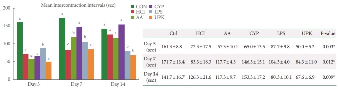
Five IC models were generated in 8-week-old female Sprague-Dawley rats via transurethral instillation of 0.1M hydrogen chloride (HCl) or 3% acetic acid (AA), intraperitoneal injection of cyclophosphamide (CYP) or lipopolysaccharide (LPS), or subcutaneous injection of uroplakin II (UPK2). After generating the IC models, conscious cystometry was performed on days 3, 7, and 14. All rats were euthanized on day 14 and their bladders were obtained for histological and pro-inflammatory-related gene expression analysis.
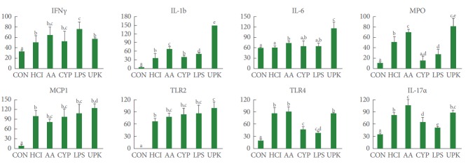
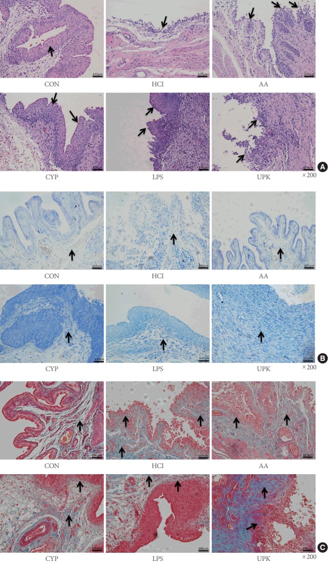
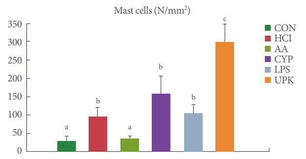
In the cystometric analysis, all experimental groups showed significantly decreased intercontraction intervals compared with the control group on day 3, but only the LPS and UPK groups maintained significantly shorter intercontraction intervals than the control group on day 14. The histological analysis revealed that areas with severe urothelial erosion (HCl, AA, and UPK) and hyperplasia (CYP and LPS), particularly in the UPK-treated bladders, showed a markedly increased infiltration of toluidine blue-stained mast cells and increased tissue fibrosis. In addition, significantly elevated expression of interleukin-1b, interleukin-6, myeloperoxidase, monocyte chemotactic protein 1, and Toll-like receptors 2 and 4 was observed in the UPK group compared to the other groups.
Among the 5 different agents, the injection of UPK generated the most effective IC animal model, showing consequent urothelial barrier loss, inflammatory reaction, tissue fibrosis stimulation, and persistent hyperactive bladder.
我们使用不同试剂评估了5种不同的大鼠模型,以便根据膀胱的功能和病理特征建立间质性膀胱炎(IC)的标准动物模型。
通过经尿道灌注0.1M盐酸(HCl)或3%乙酸(AA)、腹腔注射环磷酰胺(CYP)或脂多糖(LPS)、或皮下注射uroplakin II(UPK2),在8周龄雌性Sprague-Dawley大鼠中建立5种IC模型。建立IC模型后,在第3、7和14天进行清醒膀胱测压。所有大鼠在第14天安乐死,获取其膀胱用于组织学和促炎相关基因表达分析。
在膀胱测压分析中,所有实验组在第3天与对照组相比,收缩间期均显著缩短,但只有LPS组和UPK组在第14天收缩间期仍显著短于对照组。组织学分析显示,存在严重尿路上皮糜烂的区域(HCl、AA和UPK组)以及增生区域(CYP和LPS组),特别是在UPK处理的膀胱中,甲苯胺蓝染色的肥大细胞浸润明显增加,组织纤维化增加。此外,与其他组相比,UPK组中白细胞介素-1β、白细胞介素-6、髓过氧化物酶、单核细胞趋化蛋白1以及Toll样受体2和4的表达显著升高。
在这5种不同试剂中,注射UPK产生了最有效的IC动物模型,表现为随后的尿路上皮屏障丧失、炎症反应、组织纤维化刺激以及持续性膀胱过度活动。