Dwivedi Chirayu, Gokhale Sandeep, Khim Hyun Gon, Oh Jeon Keon, Shon Won Yong
Department of Orthopaedic Surgery, Korea University Guro Hospital, Seoul, Korea.
Hip Pelvis. 2017 Sep;29(3):168-175. doi: 10.5371/hp.2017.29.3.168. Epub 2017 Sep 6.
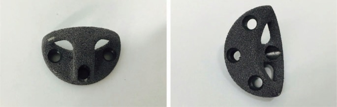
High rates of mechanical failure have been reported in type III acetabular defects. Recently porous trabecular metal augments have been introduced with, excellent biomechanical characteristics and biocompatibility, allowing early stability and greater bone ingrowth. The aim of the study was to assess the short term clinical and radiological outcome of the trabecular metal augments.
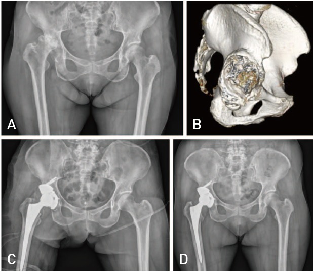
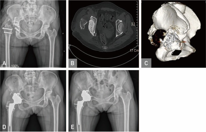
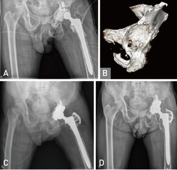
We performed, 22 revision total hip arthroplasties (THA) and 6 primary THA (total 28) using trabecular metal augments to reconstruct acetabular defect between 2011 to 2015. Among 28 patients, 18 were males, 10 females. Mean age of patients was 61.21 years. Paprosky classification for acetabular bone defects was used. Eighteen cases were classified as grade 3 A and 10 cases as grade 3B. Hip center was calculated in each case preoperatively and compared postoperatively to check whether it has been brought down. Clinical outcome assessed using Harris hip score (HHS) and radiological outcomes as osteolysis in acetabular zones and osseointegration, according to Moore's criteria.
HHS improved from 58.00 to 86.20. Centre of rotation of hip joint corrected from 38.90 mm preoperatively to 23.85 mm postoperatively above the interteardrop line. Among 28 patients, 18 patients had three or more signs of osseointegration (Moore's criteria), during final follow up and 10 had one/two signs. No radiolucency, osteolysis, or loosening found during follow up radiographic examination.
Our study showed that trabecular metal augments were highly satisfactory in short term. However, long term study is required for better evaluation.
据报道,Ⅲ型髋臼缺损的机械故障率较高。近年来,多孔小梁金属增强物被引入,具有优异的生物力学特性和生物相容性,可实现早期稳定性并促进更多骨长入。本研究的目的是评估小梁金属增强物的短期临床和放射学结果。
2011年至2015年期间,我们使用小梁金属增强物进行了22例翻修全髋关节置换术(THA)和6例初次THA(共28例),以重建髋臼缺损。28例患者中,男性18例,女性10例。患者平均年龄为61.21岁。采用髋臼骨缺损的Paprosky分类法。18例被分类为3A级,10例为3B级。术前计算每例患者的髋关节中心,并在术后进行比较,以检查其是否下移。使用Harris髋关节评分(HHS)评估临床结果,并根据Moore标准评估髋臼区域的骨溶解和骨整合等放射学结果。
HHS从58.00提高到86.20。髋关节旋转中心从术前泪滴线以上38.90 mm校正至术后的23.85 mm。28例患者中,18例在末次随访时有三个或更多骨整合迹象(Moore标准),10例有一个/两个迹象。随访X线检查未发现透光线、骨溶解或松动。
我们的研究表明,小梁金属增强物在短期内效果非常令人满意。然而,需要进行长期研究以进行更好的评估。