Wu Kun-Wei, Mo Jia-Lin, Kou Zeng-Wei, Liu Qi, Lv Ling-Ling, Lei Yu, Sun Feng-Yan
Institute of Biomedical Sciences and Department of Neurobiology, School of Basic Medical Sciences, Shanghai Medical College, Fudan UniversityShanghai, China.
Shanghai Key Laboratory of Clinical Geriatric Medicine, Research Center on Aging and Medicine, Shanghai Medical College, Fudan UniversityShanghai, China.
Front Cell Neurosci. 2017 Sep 15;11:290. doi: 10.3389/fncel.2017.00290. eCollection 2017.
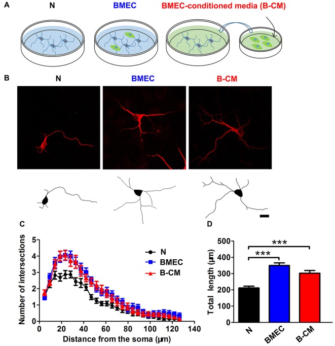
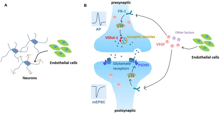
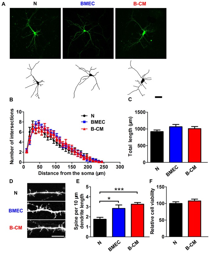
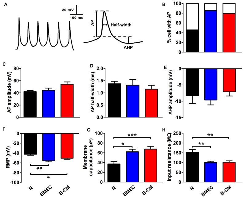
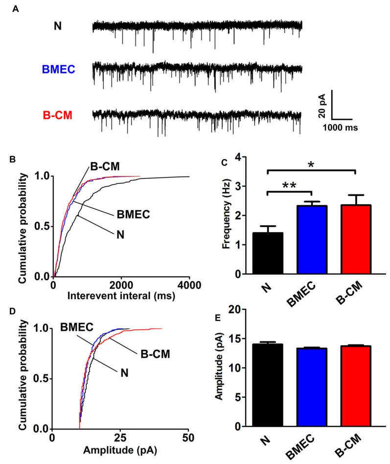
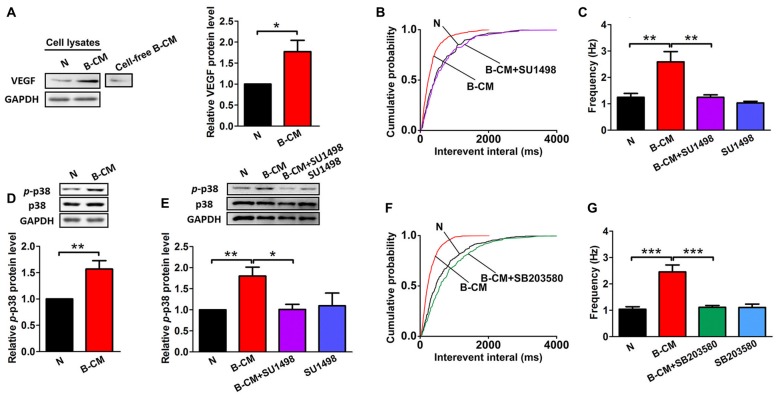
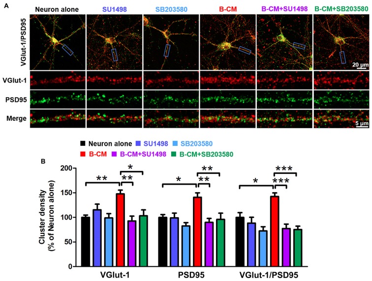
Brain microvascular endothelial cells (BMEC) have been found to guide the migration, promote the survival and regulate the differentiation of neural cells. However, whether BMEC promote development and maturation of immature neurons is still unknown. Therefore, in this study, we used a direct endothelium-neuron co-culture system combined with patch clamp recordings and confocal imaging analysis, to investigate the effects of endothelial cells on neuronal morphology and function during development. We found that endothelial cells co-culture or BMEC-conditioned medium (B-CM) promoted neurite outgrowth and spine formation, accelerated electrophysiological development and enhanced synapse function. Moreover, B-CM treatment induced vascular endothelial growth factor (VEGF) expression and p38 phosphorylation in the cortical neurons. Through pharmacological analysis, we found that incubation with SU1498, an inhibitor of VEGF receptor, abolished B-CM-induced -p38 upregulation and suppressed the enhancement of synapse formation and transmission. SB203580, an inhibitor of p38 MAPK also blocked B-CM-mediated synaptic regulation. Together these results clearly reveal that the endothelium-neuron interactions promote morphological and functional maturation of neurons. In addition, neurovascular interaction-mediated promotion of neural network maturation relies on activation of VEGF/Flk-1/p38 MAPK signaling. This study provides novel aspects of endothelium-neuron interactions and novel mechanism of neurovascular crosstalk.
已发现脑微血管内皮细胞(BMEC)可引导神经细胞迁移、促进其存活并调节其分化。然而,BMEC是否促进未成熟神经元的发育和成熟仍不清楚。因此,在本研究中,我们使用直接的内皮细胞-神经元共培养系统,结合膜片钳记录和共聚焦成像分析,来研究发育过程中内皮细胞对神经元形态和功能的影响。我们发现内皮细胞共培养或BMEC条件培养基(B-CM)可促进神经突生长和棘突形成,加速电生理发育并增强突触功能。此外,B-CM处理可诱导皮质神经元中血管内皮生长因子(VEGF)表达和p38磷酸化。通过药理学分析,我们发现用VEGF受体抑制剂SU1498孵育可消除B-CM诱导的p38上调,并抑制突触形成和传递的增强。p38丝裂原活化蛋白激酶(MAPK)抑制剂SB203580也可阻断B-CM介导的突触调节。这些结果共同清楚地表明,内皮细胞-神经元相互作用可促进神经元的形态和功能成熟。此外,神经血管相互作用介导的神经网络成熟促进依赖于VEGF/Flk-1/p38 MAPK信号通路的激活。本研究揭示了内皮细胞-神经元相互作用的新方面以及神经血管串扰的新机制。