Soltys John, Wang Qin, Mao-Draayer Yang
Present Address: University of Colorado Medical Scientist Training Program (MSTP), Aurora, CO, USA.
Department of Neurology, University of Michigan Medical School, Ann Arbor, MI, USA.
Neural Regen Res. 2017 Aug;12(8):1352-1356. doi: 10.4103/1673-5374.213558.
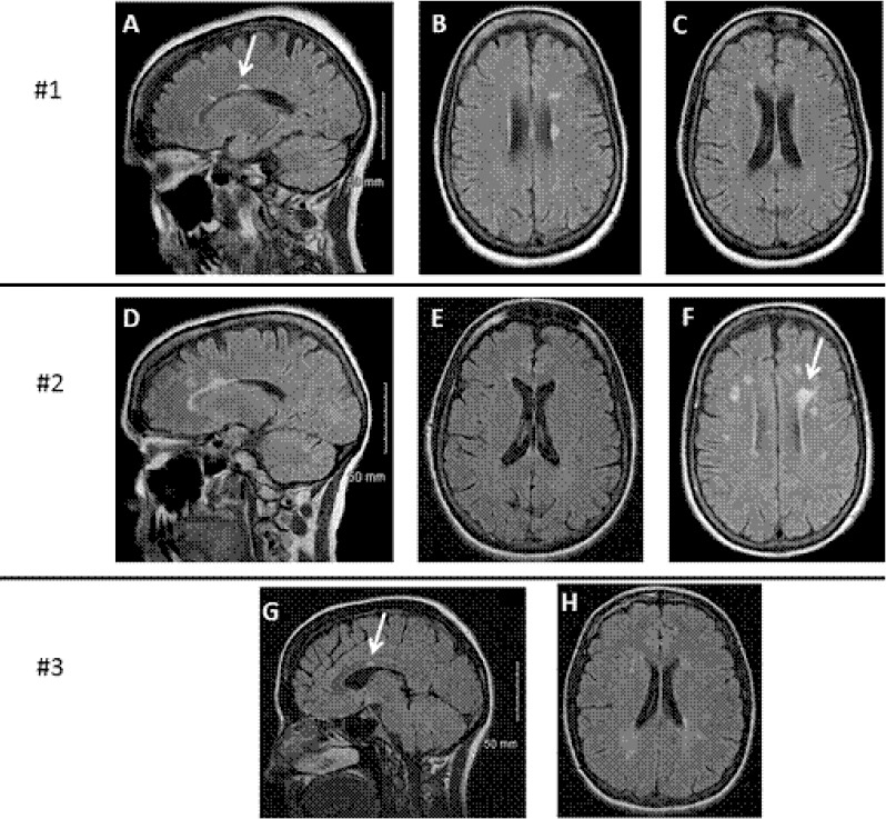
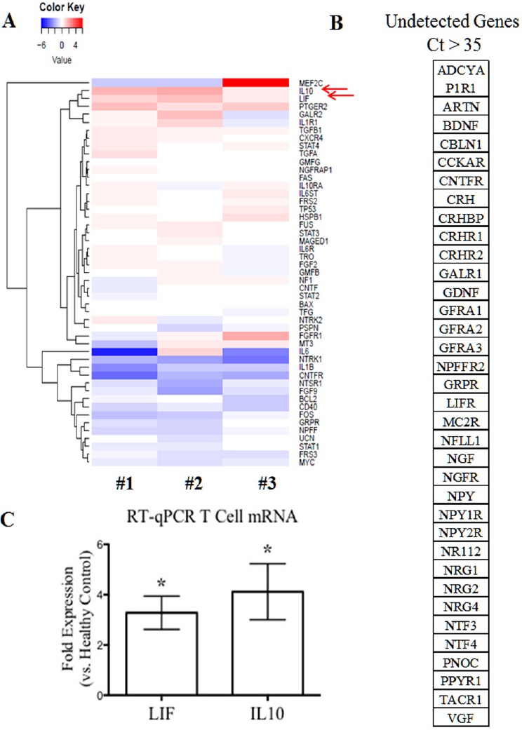
Benign multiple sclerosis is a retrospective diagnosis based primarily on a lack of motor symptom progression. Recent findings that suggest patients with benign multiple sclerosis experience non-motor symptoms highlight the need for a more prospective means to diagnose benign multiple sclerosis early in order to help direct patient care. In this study, we present optical coherence tomography and T cell neurotrophin gene analysis findings in a small number of patients with benign multiple sclerosis. Our results demonstrated that retinal nerve fiber layer was mildly thinned, and T cells had a distinct gene expression profile that included upregulation of interleukin 10 and leukemia inhibitory factor, downregulation of interleukin 6 and neurotensin high affinity receptor 1 (a novel neurotrophin receptor). These findings add evidence for further investigation into optical coherence tomography and mRNA profiling in larger cohorts as a potential means to diagnose benign multiple sclerosis in a more prospective manner.
良性多发性硬化症是一种主要基于运动症状无进展的回顾性诊断。近期研究结果表明,患有良性多发性硬化症的患者会出现非运动症状,这凸显了采用更具前瞻性的方法以便在早期诊断良性多发性硬化症从而指导患者护理的必要性。在本研究中,我们展示了少数良性多发性硬化症患者的光学相干断层扫描和T细胞神经营养因子基因分析结果。我们的结果表明,视网膜神经纤维层轻度变薄,并且T细胞具有独特的基因表达谱,包括白细胞介素10和白血病抑制因子上调,白细胞介素6和神经降压素高亲和力受体1(一种新型神经营养因子受体)下调。这些发现为在更大队列中进一步研究光学相干断层扫描和mRNA谱分析提供了证据,作为以更具前瞻性的方式诊断良性多发性硬化症的潜在手段。