Iorga Raluca Eugenia, Moraru Andreea, Ozturk Manuela Ramona, Costin Dănuţ
Department of Ophthalmology, "N. Oblu" Clinical Emergency Hospital, Iaşi, Romania.
Department of Ophthalmology, "Gr. T. Popa" University of Medicine, Iaşi, Romania.
Rom J Ophthalmol. 2018 Jan-Mar;62(1):3-14.
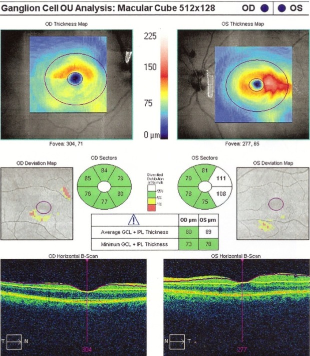
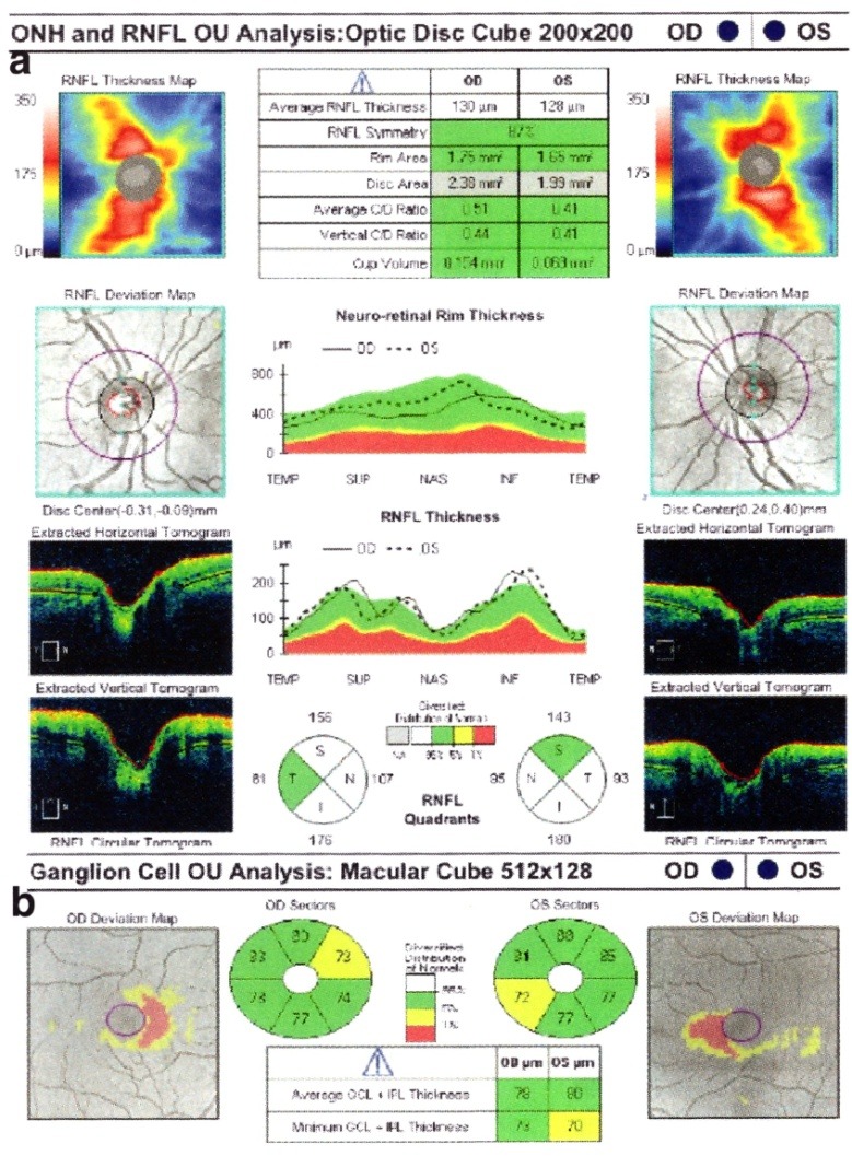
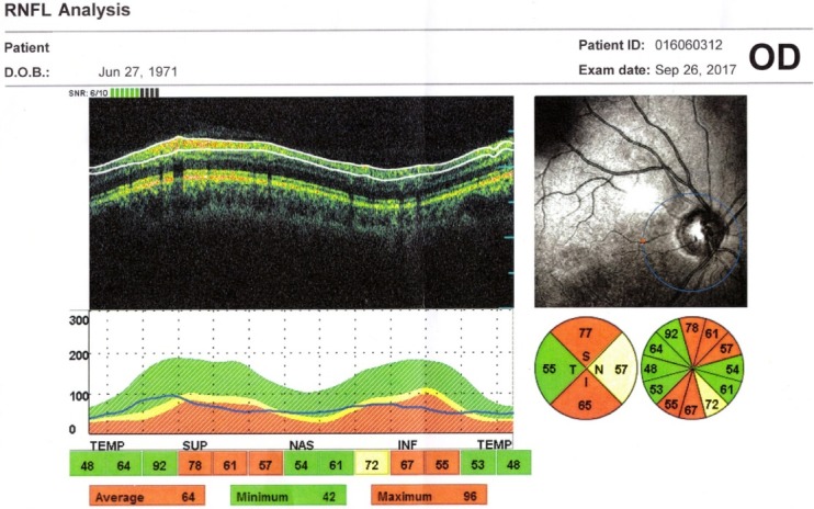
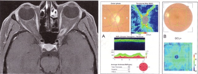
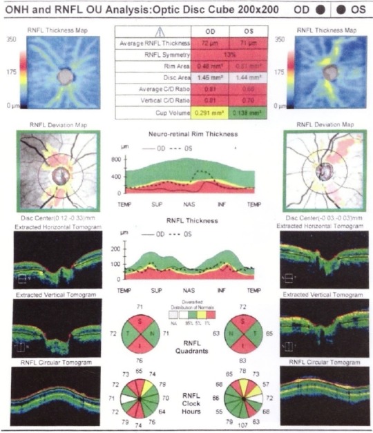
Optical neuropathies are neuro-ophthalmologic disorders, the main symptoms of which are the decrease of visual acuity and the alteration of the color vision. Optical coherence tomography has been one of the most important innovations in ophthalmology, which offered the possibility to analyze specific structures of the retina. Optical coherence tomography performs in vivo, real-time, noncontact scanning and provides cross-sectional and volumetric images with a resolution approaching that of histology. Optical coherence tomography offers the opportunity to study neurological diseases in an objective and non-invasive manner. The measurements of retinal nerve fiber layer can be an objective measurement of nerve swelling or nerve atrophy. By analyzing the ganglion cell complex, optical coherence tomography can help detect early axonal damage and may predict the visual outcome. It can be useful for diagnosis and follow-up of optic nerve and chiasmal compressive diseases. Furthermore, optical coherence tomography is useful in patients with multiple sclerosis in distinguishing macular disease from optic neuritis and in monitoring the treatment. Multiple studies and clinical observations support the importance of optical coherence tomography in the diagnosis, treatment, and follow-up of optic neuropathies.
OCT = optical coherence tomography, VA = visual acuity, RNFL = retinal nerve fiber layer, GCL = ganglion cells layer, MS = multiple sclerosis, ON = optic neuropathy, NAION = non-arteritic ischemic anterior optic neuropathy, LHON = Leber hereditary optic neuropathy, RE = right eye, LE = left eye.
视神经病变是神经眼科疾病,其主要症状是视力下降和色觉改变。光学相干断层扫描是眼科最重要的创新之一,它为分析视网膜的特定结构提供了可能。光学相干断层扫描可进行活体、实时、非接触式扫描,并提供分辨率接近组织学的横断面和容积图像。光学相干断层扫描为以客观和非侵入性方式研究神经系统疾病提供了机会。视网膜神经纤维层的测量可以是神经肿胀或神经萎缩的客观测量方法。通过分析神经节细胞复合体,光学相干断层扫描有助于检测早期轴突损伤,并可能预测视力预后。它对视神经和视交叉压迫性疾病的诊断和随访可能有用。此外,光学相干断层扫描在多发性硬化症患者中有助于区分黄斑疾病和视神经炎,并监测治疗情况。多项研究和临床观察支持光学相干断层扫描在视神经病变的诊断、治疗和随访中的重要性。
OCT = 光学相干断层扫描,VA = 视力,RNFL = 视网膜神经纤维层,GCL = 神经节细胞层,MS = 多发性硬化症,ON = 视神经病变,NAION = 非动脉炎性前部缺血性视神经病变,LHON = 莱伯遗传性视神经病变,RE = 右眼,LE = 左眼。