1 Department of Cardiovascular Surgery, Osaka University Graduate School of Medicine , Suita, Osaka, Japan .
2 Department of Applied Chemistry, Osaka University Graduate School of Engineering , Osaka, Japan .
Tissue Eng Part C Methods. 2018 Jan;24(1):56-67. doi: 10.1089/ten.TEC.2017.0247. Epub 2017 Nov 17.
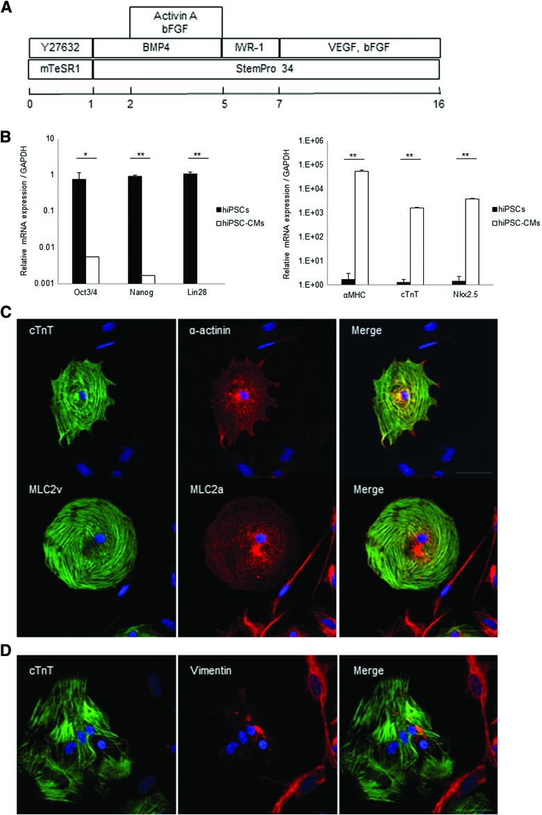
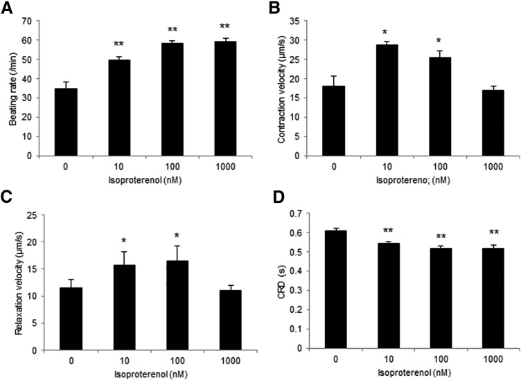
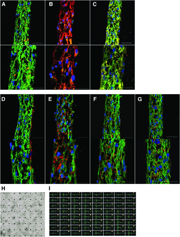
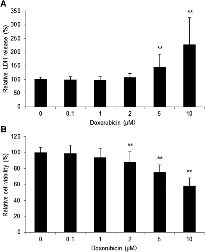
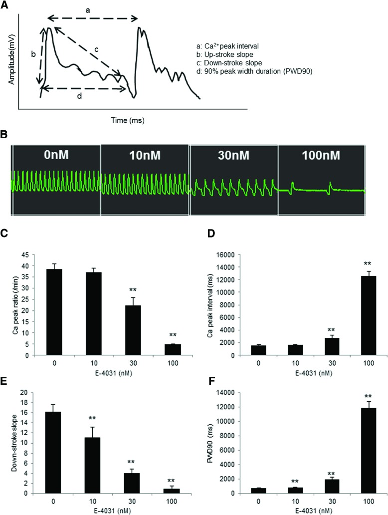
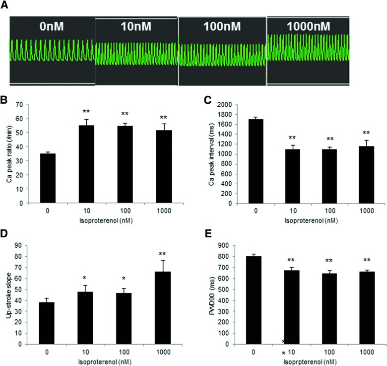
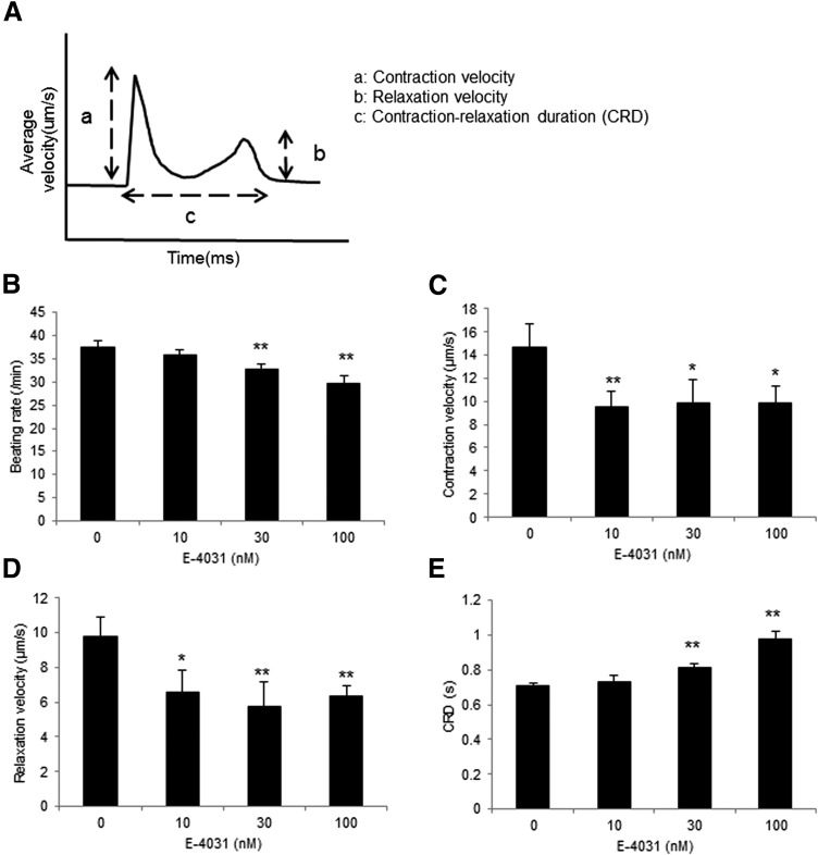
An in vitro drug-induced cardiotoxicity assay is a critical step in drug discovery for clinical use. The use of human induced pluripotent stem cell-derived cardiomyocytes (hiPSC-CMs) is promising for this purpose. However, single hiPSC-CMs are limited in their ability to mimic native cardiac tissue structurally and functionally, and the generation of artificial cardiac tissue using hiPSC-CMs is an ongoing challenging. We therefore developed a new method of constructing three-dimensional (3D) artificial tissues in a short time by coating extracellular matrix (ECM) components on cell surfaces. We hypothesized that 3D cardiac tissues derived from hiPSC-CMs (3D-hiPSC-CT) could be used for an in vitro drug-induced cardiotoxicity assay. 3D-hiPSC-CT were generated by fibronectin and gelatin nanofilm coated single hiPSC-CMs. Histologically, 3D-hiPSC-CT exhibited a sarcomere structure in the myocytes and ECM proteins, such as fibronectin, collagen type I/III, and laminin. The administration of cytotoxic doxorubicin at 5.0 μM induced the release of lactate dehydrogenase, while that at 2.0 μM reduced the cell viability. E-4031, human ether-a-go-go related gene (hERG)-type potassium channel blocker, and isoproterenol induced significant changes both in the Ca transient parameters and contractile parameters in a dose-dependent manner. The 3D-hiPSC-CT exhibited doxorubicin-sensitive cytotoxicity and hERG channel blocker/isoproterenol-sensitive electrical activity in vitro, indicating its usefulness for drug-induced cardiotoxicity assays or drug screening systems for drug discovery.
体外药物诱导的心脏毒性检测是临床应用药物发现的关键步骤。人诱导多能干细胞衍生的心肌细胞(hiPSC-CMs)在这方面很有前途。然而,单个 hiPSC-CMs 在结构和功能上模拟天然心脏组织的能力有限,并且使用 hiPSC-CMs 生成人工心脏组织仍然具有挑战性。因此,我们开发了一种新的方法,通过在细胞表面涂覆细胞外基质(ECM)成分,在短时间内构建三维(3D)人工组织。我们假设,来自 hiPSC-CMs 的 3D 心脏组织(3D-hiPSC-CT)可用于体外药物诱导的心脏毒性检测。3D-hiPSC-CT 通过纤连蛋白和明胶纳米膜涂覆的单个 hiPSC-CMs 生成。组织学上,3D-hiPSC-CT 在心肌细胞和 ECM 蛋白(如纤连蛋白、I/III 型胶原蛋白和层粘连蛋白)中表现出肌节结构。给予细胞毒性阿霉素 5.0μM 可诱导乳酸脱氢酶释放,而给予 2.0μM 则降低细胞活力。E-4031(人 Ether-a-go-go 相关基因(hERG)型钾通道阻滞剂)和异丙肾上腺素以剂量依赖性方式诱导钙瞬变参数和收缩参数的显著变化。3D-hiPSC-CT 在体外表现出对阿霉素的敏感性细胞毒性和 hERG 通道阻滞剂/异丙肾上腺素的敏感性电活性,表明其在药物诱导的心脏毒性检测或药物发现的药物筛选系统中具有用途。