Translational Neuroradiology Section, National Institute of Neurological Disorders and Stroke, National Institutes of Health, Bethesda, United States.
Hematopathology Section, Laboratory of Pathology, National Cancer Institute, National Institutes of Health, Bethesda, United States.
Elife. 2017 Oct 3;6:e29738. doi: 10.7554/eLife.29738.
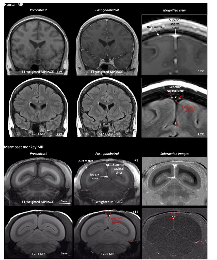
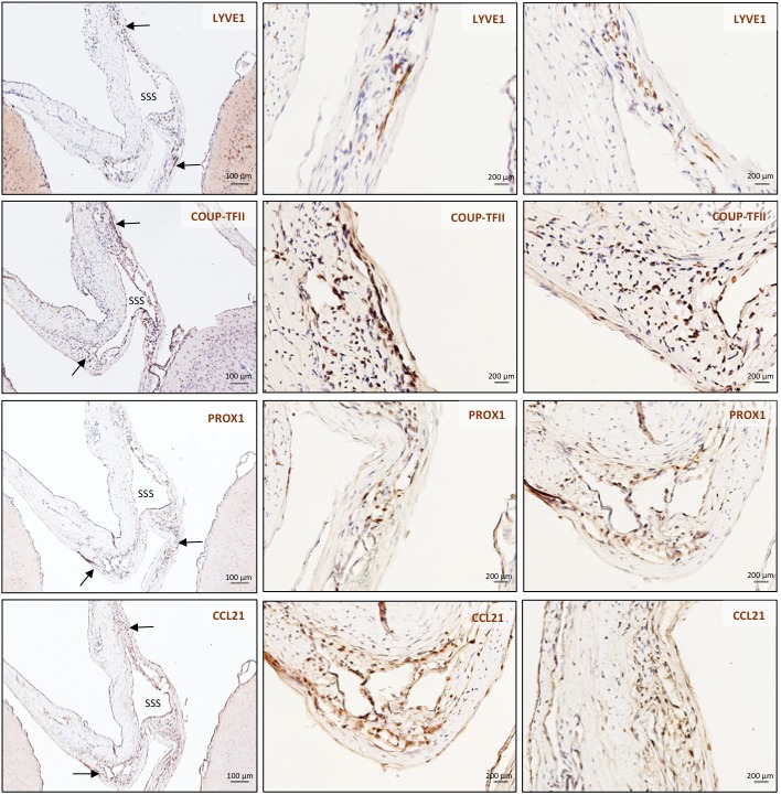
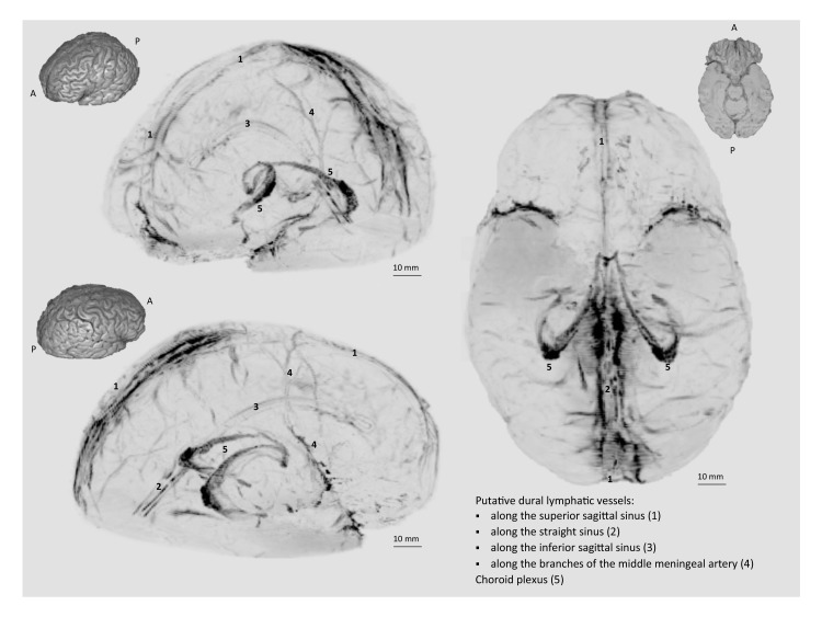
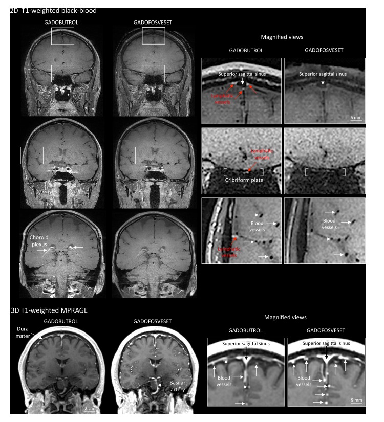
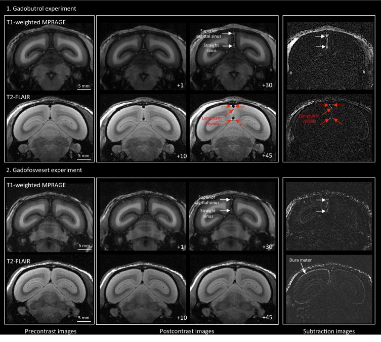
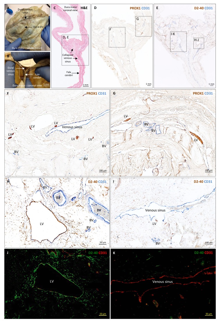
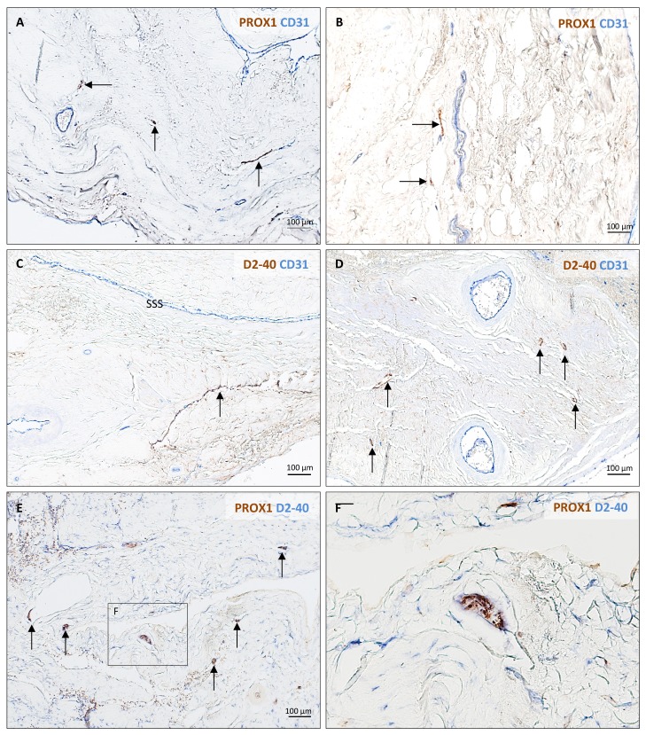
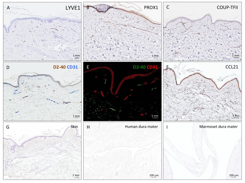
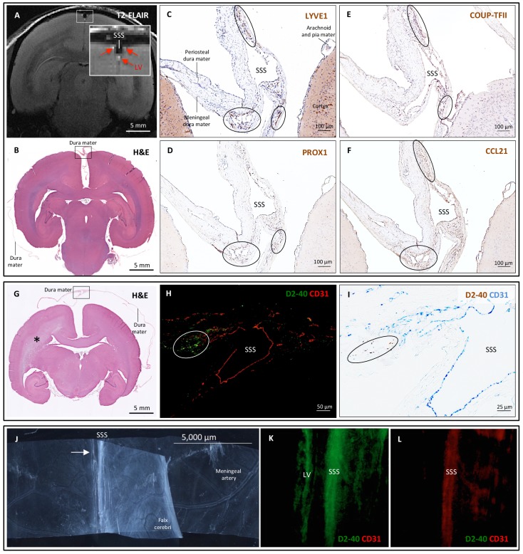
Here, we report the existence of meningeal lymphatic vessels in human and nonhuman primates (common marmoset monkeys) and the feasibility of noninvasively imaging and mapping them in vivo with high-resolution, clinical MRI. On T2-FLAIR and T1-weighted black-blood imaging, lymphatic vessels enhance with gadobutrol, a gadolinium-based contrast agent with high propensity to extravasate across a permeable capillary endothelial barrier, but not with gadofosveset, a blood-pool contrast agent. The topography of these vessels, running alongside dural venous sinuses, recapitulates the meningeal lymphatic system of rodents. In primates, meningeal lymphatics display a typical panel of lymphatic endothelial markers by immunohistochemistry. This discovery holds promise for better understanding the normal physiology of lymphatic drainage from the central nervous system and potential aberrations in neurological diseases.
在这里,我们报告了脑膜淋巴管在人类和非人类灵长类动物(普通狨猴)中的存在,以及使用高分辨率临床 MRI 对其进行非侵入性成像和体内绘图的可行性。在 T2-FLAIR 和 T1 加权黑血成像中,淋巴管增强与钆布醇增强,这是一种具有高倾向穿过可渗透的毛细血管内皮屏障外渗的基于钆的造影剂,但与血池造影剂钆氟塞特不同。这些与硬脑膜静脉窦并行运行的血管的拓扑结构再现了啮齿动物的脑膜淋巴系统。在灵长类动物中,脑膜淋巴管通过免疫组织化学显示出典型的一组淋巴管内皮标志物。这一发现有望更好地理解从中枢神经系统进行淋巴引流的正常生理学以及神经疾病中的潜在异常。