Institut du Cerveau, Pitié-Salpêtrière Hospital, Centre National de la Recherche Scientifique, Institut National de la Santé et de la Recherche Médicale, Sorbonne Université, Paris, France.
Paris Cardiovascular Research Center, Institut National de la Santé et de la Recherche Médicale, Université de Paris, Paris, France.
J Exp Med. 2022 Aug 1;219(8). doi: 10.1084/jem.20220035. Epub 2022 Jul 1.
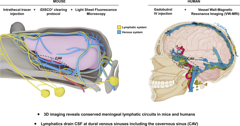
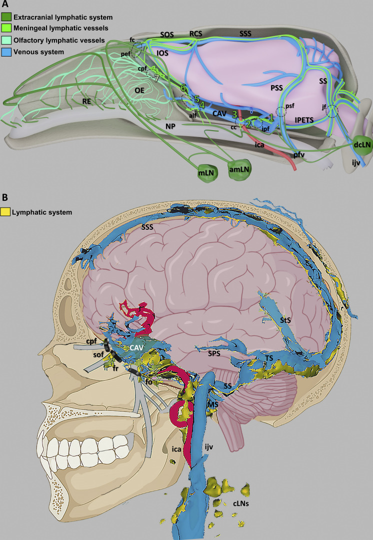
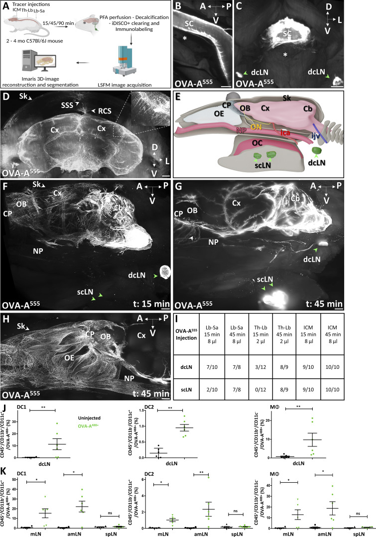
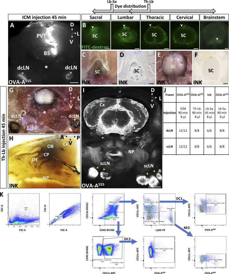
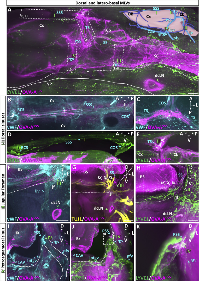
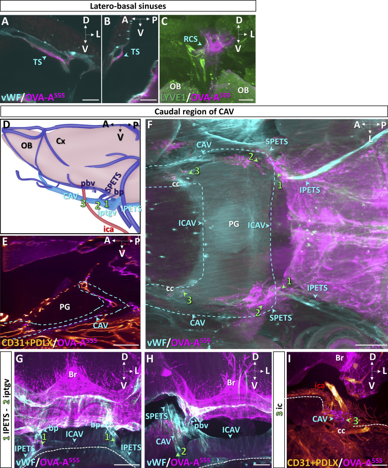
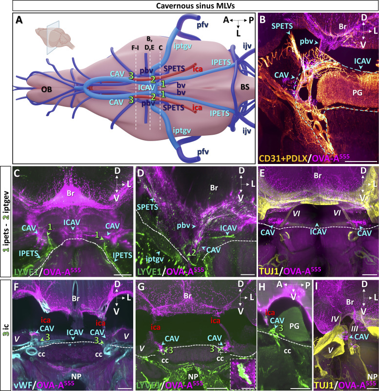
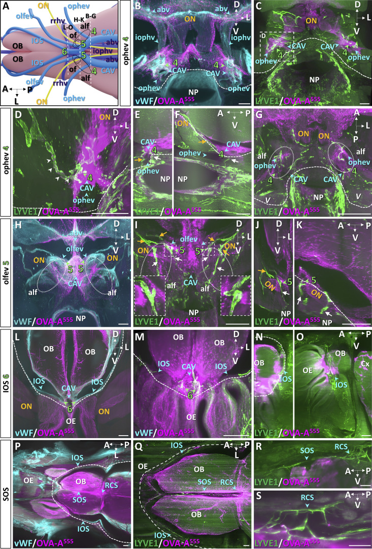
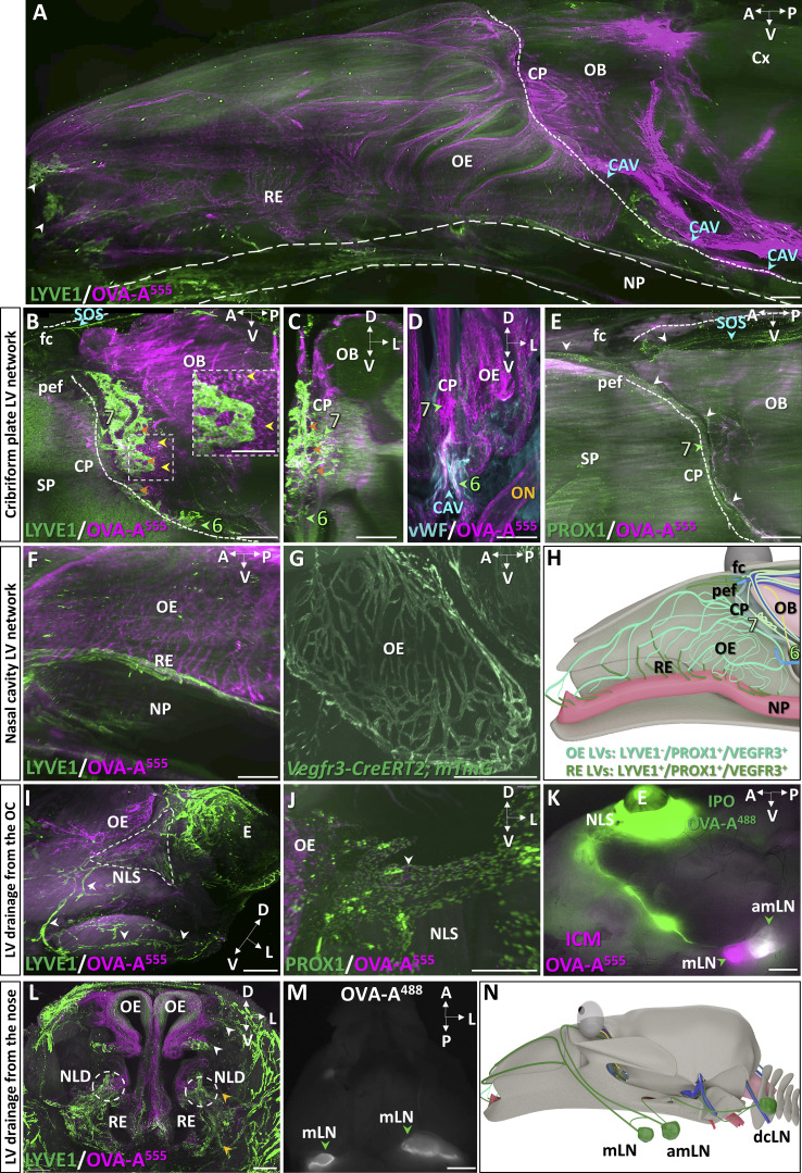
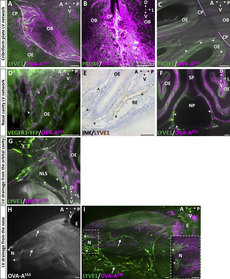
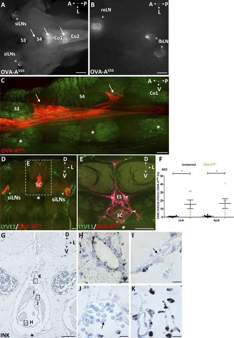
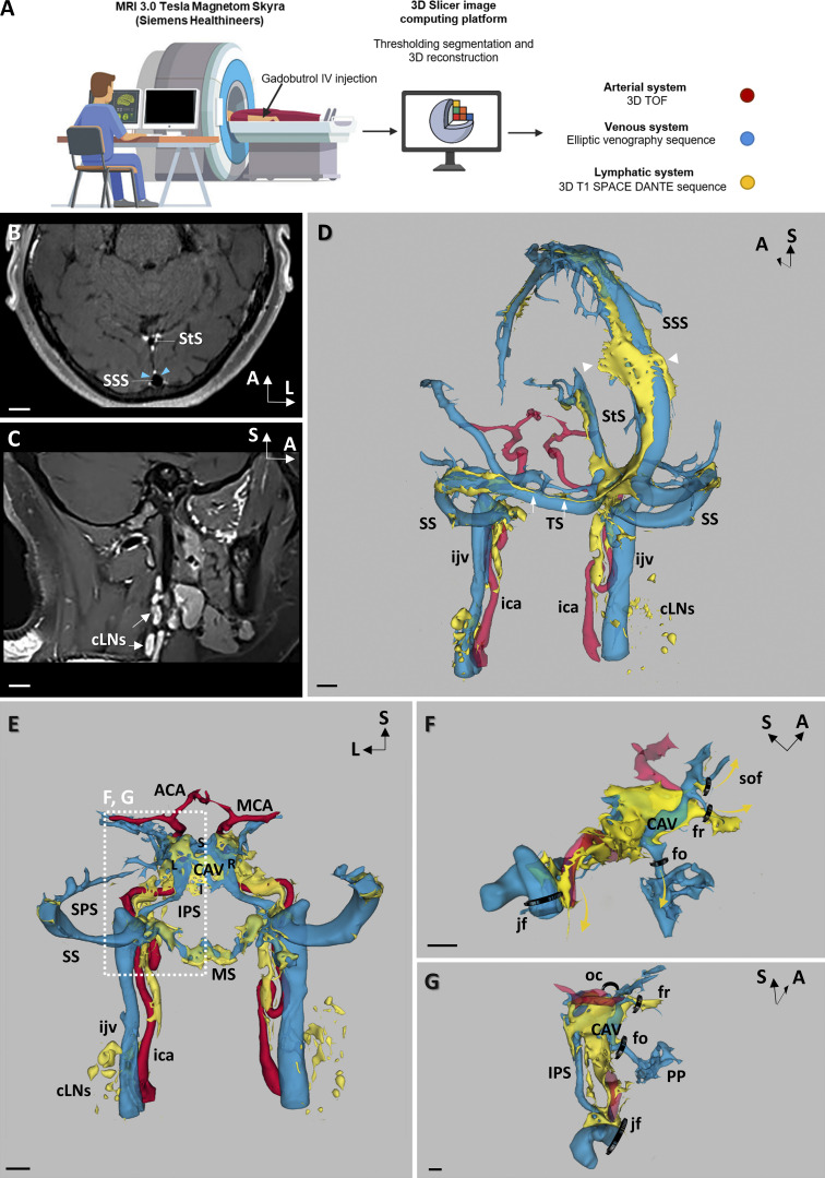
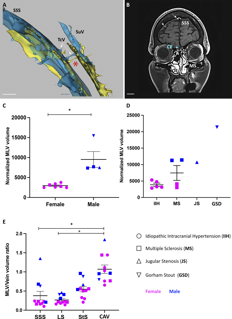
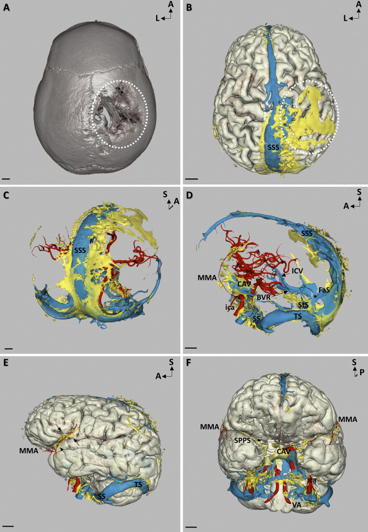
Meningeal lymphatic vessels (MLVs) were identified in the dorsal and caudobasal regions of the dura mater, where they ensure waste product elimination and immune surveillance of brain tissues. Whether MLVs exist in the anterior part of the murine and human skull and how they connect with the glymphatic system and extracranial lymphatics remained unclear. Here, we used light-sheet fluorescence microscopy (LSFM) imaging of mouse whole-head preparations after OVA-A555 tracer injection into the cerebrospinal fluid (CSF) and performed real-time vessel-wall (VW) magnetic resonance imaging (VW-MRI) after systemic injection of gadobutrol in patients with neurological pathologies. We observed a conserved three-dimensional anatomy of MLVs in mice and humans that aligned with dural venous sinuses but not with nasal CSF outflow, and we discovered an extended anterior MLV network around the cavernous sinus, with exit routes through the foramina of emissary veins. VW-MRI may provide a diagnostic tool for patients with CSF drainage defects and neurological diseases.
脑膜淋巴管 (MLVs) 被发现在硬脑膜的背侧和尾基底区域,它们确保了脑组织的废物清除和免疫监视。MLVs 是否存在于鼠类和人类颅骨的前部,以及它们如何与神经胶淋巴系统和颅外淋巴管相连,这些仍然不清楚。在这里,我们使用 OVA-A555 示踪剂注入脑脊液 (CSF) 后的小鼠全脑标本的光片荧光显微镜 (LSFM) 成像,并在患有神经病理学的患者中进行了全身性注射钆布醇后的血管壁 (VW) 磁共振成像 (VW-MRI)。我们观察到了在小鼠和人类中保守的 MLV 的三维解剖结构,它们与硬脑膜静脉窦一致,但与鼻 CSF 流出无关,并且我们发现了一个围绕海绵窦的扩展的前 MLV 网络,其通过导血管的孔道离开。VW-MRI 可能为 CSF 引流缺陷和神经疾病患者提供一种诊断工具。