• 文献检索
  • 文档翻译
  • 深度研究
  • 学术资讯
  • Suppr Zotero 插件Zotero 插件
  • 邀请有礼
  • 套餐&价格
  • 历史记录
应用&插件
Suppr Zotero 插件Zotero 插件浏览器插件Mac 客户端Windows 客户端微信小程序
定价
高级版会员购买积分包购买API积分包
服务
文献检索文档翻译深度研究API 文档MCP 服务
关于我们
关于 Suppr公司介绍联系我们用户协议隐私条款
关注我们

Suppr 超能文献

核心技术专利:CN118964589B侵权必究
粤ICP备2023148730 号-1Suppr @ 2026

文献检索

告别复杂PubMed语法,用中文像聊天一样搜索,搜遍4000万医学文献。AI智能推荐,让科研检索更轻松。

立即免费搜索

文件翻译

保留排版,准确专业,支持PDF/Word/PPT等文件格式,支持 12+语言互译。

免费翻译文档

深度研究

AI帮你快速写综述,25分钟生成高质量综述,智能提取关键信息,辅助科研写作。

立即免费体验

淋巴样增强因子-1:在鉴别涎腺基底样肿瘤中的诊断效用

LEF-1: Diagnostic utility in distinguishing basaloid neoplasms of the salivary gland.

作者信息

Schmitt Alessandra C, Griffith Christopher C, Cohen Cynthia, Siddiqui Momin T

机构信息

Emory University School of Medicine, Pathology, Atlanta, Georgia.

出版信息

Diagn Cytopathol. 2017 Dec;45(12):1078-1083. doi: 10.1002/dc.23820. Epub 2017 Oct 3.

DOI:10.1002/dc.23820
PMID:28972308
原文链接:https://pmc.ncbi.nlm.nih.gov/articles/PMC5930010/
Abstract

BACKGROUND

Lymphoid enhancer binding factor 1 (LEF-1) has recently been reported as a potential immunohistochemical (IHC) marker for basal cell adenoma (BCA) and other salivary gland tumors, which may contribute to an increased accuracy in differentiating basaloid salivary gland neoplasms. We evaluated the utility of LEF-1 in fine needle aspiration (FNA) and resection specimens to distinguish pleomorphic adenoma (PA), BCA, basal cell adenocarcinoma (BCAC), and adenoid cystic carcinoma (ACC) as well as in non-neoplastic salivary gland (NNSG).

METHODS

Cases including 66 PA (35 FNA, 31 resections), 12 BCA (5 FNA, 7 resections), 42 ACC (11 FNA, 31 resections), 1 BCAC FNA, and 10 NNSG (5 FNA, 5 resections) were obtained and stained for LEF-1.

RESULTS

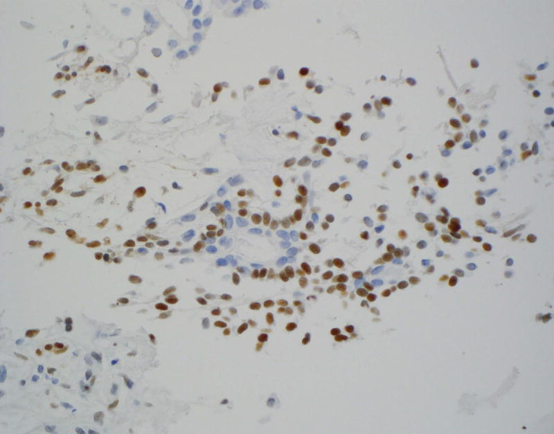
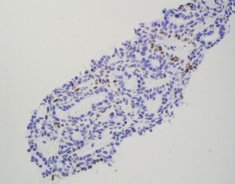
On cell block (CB), 51% of PA and 60% of BCA were LEF-1 positive while 91% of ACC were LEF-1 negative. Among resections, there was a higher percentage of LEF-1 positive PA (84%) and BCA (86%), and a higher percentage of LEF-1 negative ACC (97%). LEF-1 staining had a low to moderate sensitivity for detecting benign basaloid neoplasms on FNA CB and resection specimens (52.5% and 84%, respectively), but a higher specificity (92% and 97% respectively), and positive predictive value (95% and 97% respectively).

CONCLUSION

When comparing benign (PA and BCA) and the most common malignant basaloid salivary gland tumor (ACC), positive LEF-1 favors a benign neoplasm. Additional studies with LEF-1, specifically including other rare basaloid salivary gland neoplasms are needed to further clarify the role of LEF-1 in diagnosing these lesions on FNA.

摘要

背景

淋巴样增强子结合因子1(LEF-1)最近被报道为基底细胞腺瘤(BCA)和其他涎腺肿瘤潜在的免疫组织化学(IHC)标志物,这可能有助于提高鉴别涎腺基底样肿瘤的准确性。我们评估了LEF-1在细针穿刺(FNA)标本和手术切除标本中鉴别多形性腺瘤(PA)、BCA、基底细胞腺癌(BCAC)和腺样囊性癌(ACC)以及在非肿瘤性涎腺(NNSG)中的应用价值。

方法

获取了包括66例PA(35例FNA,31例手术切除)、12例BCA(5例FNA,7例手术切除)、42例ACC(11例FNA,31例手术切除)、1例BCAC FNA以及10例NNSG(5例FNA,5例手术切除)的病例,并进行LEF-1染色。

结果

在细胞块(CB)上,51%的PA和60%的BCA为LEF-1阳性,而91%的ACC为LEF-1阴性。在手术切除标本中,LEF-1阳性的PA(84%)和BCA(86%)比例更高,LEF-1阴性的ACC(97%)比例更高。LEF-1染色在FNA CB和手术切除标本上检测良性基底样肿瘤的敏感性较低至中等(分别为52.5%和84%),但特异性较高(分别为92%和97%),阳性预测值也较高(分别为95%和97%)。

结论

在比较良性(PA和BCA)和最常见的恶性涎腺基底样肿瘤(ACC)时,LEF-1阳性支持良性肿瘤。需要对LEF-进行更多研究,特别是纳入其他罕见的涎腺基底样肿瘤,以进一步阐明LEF-1在FNA诊断这些病变中的作用。

https://cdn.ncbi.nlm.nih.gov/pmc/blobs/5cff/5930010/b11170fc8c5f/nihms929069f2a.jpg
https://cdn.ncbi.nlm.nih.gov/pmc/blobs/5cff/5930010/20174bf32842/nihms929069f1a.jpg
https://cdn.ncbi.nlm.nih.gov/pmc/blobs/5cff/5930010/b11170fc8c5f/nihms929069f2a.jpg
https://cdn.ncbi.nlm.nih.gov/pmc/blobs/5cff/5930010/20174bf32842/nihms929069f1a.jpg
https://cdn.ncbi.nlm.nih.gov/pmc/blobs/5cff/5930010/b11170fc8c5f/nihms929069f2a.jpg

相似文献

1
LEF-1: Diagnostic utility in distinguishing basaloid neoplasms of the salivary gland.淋巴样增强因子-1:在鉴别涎腺基底样肿瘤中的诊断效用
Diagn Cytopathol. 2017 Dec;45(12):1078-1083. doi: 10.1002/dc.23820. Epub 2017 Oct 3.
2
Wnt/β-catenin signal alteration and its diagnostic utility in basal cell adenoma and histologically similar tumors of the salivary gland.Wnt/β-连环蛋白信号改变及其在涎腺基底细胞腺瘤和组织学相似肿瘤中的诊断应用
Pathol Res Pract. 2018 Apr;214(4):586-592. doi: 10.1016/j.prp.2017.12.016. Epub 2018 Jan 3.
3
PLAG1: An Immunohistochemical Marker with Limited Utility in Separating Pleomorphic Adenoma from Other Basaloid Salivary Gland Tumors.PLAG1:一种在鉴别多形性腺瘤与其他基底样唾液腺肿瘤方面效用有限的免疫组织化学标志物。
Acta Cytol. 2016;60(3):240-5. doi: 10.1159/000447622. Epub 2016 Jul 28.
4
MYB expression: Potential role in separating adenoid cystic carcinoma (ACC) from pleomorphic adenoma (PA).MYB表达:在鉴别腺样囊性癌(ACC)与多形性腺瘤(PA)中的潜在作用。
Diagn Cytopathol. 2016 Oct;44(10):799-804. doi: 10.1002/dc.23551. Epub 2016 Aug 5.
5
Pitfalls of fine-needle aspiration cytology of parotid membranous basal cell adenoma-A review of pitfalls in FNA cytology of salivary gland neoplasms with basaloid cell features.腮腺膜性基底细胞腺瘤细针穿刺细胞学检查的陷阱——对具有基底样细胞特征的唾液腺肿瘤细针穿刺细胞学检查陷阱的综述
Diagn Cytopathol. 2015 May;43(5):432-7. doi: 10.1002/dc.23234. Epub 2014 Nov 27.
6
Adenoid cystic carcinoma cytology: Salivary gland and nonsalivary gland.腺样囊性癌细胞学:唾液腺和非唾液腺。
Diagn Cytopathol. 2020 Dec;48(12):1282-1289. doi: 10.1002/dc.24573. Epub 2020 Aug 18.
7
The PRKD1 E710D hotspot mutation is highly specific in separating polymorphous adenocarcinoma of the palate from adenoid cystic carcinoma and pleomorphic adenoma on FNA.PRKD1 E710D 热点突变在 FNA 中对分离腭部多形性腺癌、腺样囊性癌和多形性腺瘤具有高度特异性。
Cancer Cytopathol. 2018 Apr;126(4):275-281. doi: 10.1002/cncy.21959. Epub 2017 Dec 20.
8
Usefulness of translocation-associated immunohistochemical stains in the fine-needle aspiration diagnosis of salivary gland neoplasms.易位相关免疫组化染色在涎腺肿瘤细针穿刺诊断中的应用价值
Cancer Cytopathol. 2016 Jun;124(6):397-405. doi: 10.1002/cncy.21693. Epub 2016 Feb 16.
9
Basal cell adenoma: a diagnostic dilemma on fine needle aspiration cytology.基底细胞腺瘤:细针穿刺细胞学检查中的诊断难题。
Diagn Cytopathol. 2011 Dec;39(12):913-6. doi: 10.1002/dc.21576. Epub 2011 Mar 4.
10
A comparative analysis of LEF-1 in odontogenic and salivary tumors.牙源性肿瘤和唾液腺肿瘤中LEF-1的比较分析。
Hum Pathol. 2015 Feb;46(2):255-9. doi: 10.1016/j.humpath.2014.10.018. Epub 2014 Nov 12.

引用本文的文献

1
Lymphoid enhancer-binding factor 1 (LEF1): a reliable immunohistochemical predictive marker for WNT-activated medulloblastoma.淋巴样增强子结合因子1(LEF1):一种用于WNT激活型髓母细胞瘤的可靠免疫组化预测标志物。
Childs Nerv Syst. 2025 Jun 17;41(1):213. doi: 10.1007/s00381-025-06872-8.
2
Intraoral Pleomorphic Adenoma of the Minor Salivary Glands: A Case Series of 10 Cases With Emphasis on Histopathological Features.小唾液腺口腔内多形性腺瘤:10例病例系列报道,重点关注组织病理学特征
Case Rep Dent. 2025 Jan 13;2025:9151514. doi: 10.1155/crid/9151514. eCollection 2025.
3
Proceedings of the 2024 North American Society of Head and Neck Pathology Companion Meeting, Baltimore, MD, March 24, 2024: Navigating Ancillary Studies in Basaloid/Blue Salivary Tumors.

本文引用的文献

1
A comparative analysis of LEF-1 in odontogenic and salivary tumors.牙源性肿瘤和唾液腺肿瘤中LEF-1的比较分析。
Hum Pathol. 2015 Feb;46(2):255-9. doi: 10.1016/j.humpath.2014.10.018. Epub 2014 Nov 12.
2
Expression of LEF1 is an independent prognostic factor for patients with oral squamous cell carcinoma.LEF1的表达是口腔鳞状细胞癌患者的一个独立预后因素。
J Formos Med Assoc. 2014 Dec;113(12):934-9. doi: 10.1016/j.jfma.2013.07.012. Epub 2013 Sep 7.
3
Nuclear overexpression of lymphoid-enhancer-binding factor 1 identifies chronic lymphocytic leukemia/small lymphocytic lymphoma in small B-cell lymphomas.
2024 年北美头颈部病理学会联合会议记录,马里兰州巴尔的摩,2024 年 3 月 24 日:蓝细胞性基底细胞样唾液腺瘤的辅助检查解读。
Head Neck Pathol. 2024 Mar 25;18(1):24. doi: 10.1007/s12105-024-01626-5.
4
Re: Mucoepidermoid Carcinoma of the Parotid Gland Mimicking Pleomorphic Adenoma on Cytology: A Case Report.回复:腮腺黏液表皮样癌在细胞学上酷似多形性腺瘤:一例报告
Indian J Otolaryngol Head Neck Surg. 2023 Sep;75(3):2737-2738. doi: 10.1007/s12070-023-03806-1. Epub 2023 May 8.
5
Basal Cell Adenoma and Basal Cell Adenocarcinoma: a 50-year Experience From a Single Institution.基底细胞腺瘤和基底细胞腺癌:单一机构 50 年经验。
Head Neck Pathol. 2022 Dec;16(4):1157-1166. doi: 10.1007/s12105-022-01484-z. Epub 2022 Aug 10.
6
A 4-Year Retrospective Analysis of Salivary Gland Cytopathology Using the Milan System for Reporting Salivary Gland Cytology and Ancillary Studies.使用米兰唾液腺细胞学报告系统及辅助检查对唾液腺细胞病理学进行的4年回顾性分析
Cancers (Basel). 2019 Dec 1;11(12):1912. doi: 10.3390/cancers11121912.
7
Consistent LEF-1 and MYB Immunohistochemical Expression in Human Papillomavirus-Related Multiphenotypic Sinonasal Carcinoma: A Potential Diagnostic Pitfall.人乳头瘤病毒相关多表型鼻窦癌中LEF-1和MYB免疫组化表达的一致性:一个潜在的诊断陷阱
Head Neck Pathol. 2019 Jun;13(2):220-224. doi: 10.1007/s12105-018-0951-1. Epub 2018 Jul 19.
核过表达淋巴增强结合因子 1 可识别小 B 细胞淋巴瘤中的慢性淋巴细胞白血病/小淋巴细胞淋巴瘤。
Mod Pathol. 2011 Nov;24(11):1433-43. doi: 10.1038/modpathol.2011.103. Epub 2011 Jun 17.
4
LEF-1 expression in basal cell carcinomas.基底细胞癌中的淋巴细胞增强因子1(LEF-1)表达
Br J Dermatol. 2009 Jun;160(6):1353-6. doi: 10.1111/j.1365-2133.2009.09144.x. Epub 2009 Mar 30.
5
Wnt3a regulates Lef-1 expression during airway submucosal gland morphogenesis.Wnt3a在气道黏膜下腺形态发生过程中调节Lef-1表达。
Dev Biol. 2007 May 1;305(1):90-102. doi: 10.1016/j.ydbio.2007.01.038. Epub 2007 Feb 7.
6
Expression patterns of hair and epithelial keratins and transcription factors HOXC13, LEF1, and beta-catenin in a malignant pilomatricoma: a histological and immunohistochemical study.恶性毛母质瘤中毛发角蛋白和上皮角蛋白以及转录因子HOXC13、LEF1和β-连环蛋白的表达模式:一项组织学和免疫组织化学研究
J Cutan Pathol. 2006 Jan;33(1):1-9. doi: 10.1111/j.0303-6987.2006.00308.x.
7
Expression of enamel proteins and LEF1 in adamantinomatous craniopharyngioma: evidence for its odontogenic epithelial differentiation.成釉蛋白和LEF1在造釉细胞瘤型颅咽管瘤中的表达:其牙源性上皮分化的证据
Histopathology. 2004 Dec;45(6):573-9. doi: 10.1111/j.1365-2559.2004.02029.x.
8
Characterization of Lef-1 promoter segments that facilitate inductive developmental expression in skin.促进皮肤诱导性发育表达的Lef-1启动子片段的特征分析。
J Invest Dermatol. 2004 Aug;123(2):264-74. doi: 10.1111/j.0022-202X.2004.23201.x.
9
On the regulation of hair keratin expression: lessons from studies in pilomatricomas.关于毛发角蛋白表达的调控:来自毛母质瘤研究的经验教训。
J Invest Dermatol. 2004 May;122(5):1078-83. doi: 10.1111/j.0022-202X.2004.22513.x.
10
Submucosal gland development in the airway is controlled by lymphoid enhancer binding factor 1 (LEF1).气道中的黏膜下腺发育受淋巴样增强子结合因子1(LEF1)控制。
Development. 1999 Oct;126(20):4441-53. doi: 10.1242/dev.126.20.4441.