• 文献检索
  • 文档翻译
  • 深度研究
  • 学术资讯
  • Suppr Zotero 插件Zotero 插件
  • 邀请有礼
  • 套餐&价格
  • 历史记录
应用&插件
Suppr Zotero 插件Zotero 插件浏览器插件Mac 客户端Windows 客户端微信小程序
定价
高级版会员购买积分包购买API积分包
服务
文献检索文档翻译深度研究API 文档MCP 服务
关于我们
关于 Suppr公司介绍联系我们用户协议隐私条款
关注我们

Suppr 超能文献

核心技术专利:CN118964589B侵权必究
粤ICP备2023148730 号-1Suppr @ 2026

文献检索

告别复杂PubMed语法,用中文像聊天一样搜索,搜遍4000万医学文献。AI智能推荐,让科研检索更轻松。

立即免费搜索

文件翻译

保留排版,准确专业,支持PDF/Word/PPT等文件格式,支持 12+语言互译。

免费翻译文档

深度研究

AI帮你快速写综述,25分钟生成高质量综述,智能提取关键信息,辅助科研写作。

立即免费体验

作为视觉功能生物标志物的固定稳定性的指标和采集模式。

Metrics and Acquisition Modes for Fixation Stability as a Visual Function Biomarker.

作者信息

Schönbach Etienne M, Ibrahim Mohamed A, Kong Xiangrong, Strauss Rupert W, Muñoz Beatriz, Birch David G, Sunness Janet S, West Sheila K, Scholl Hendrik P N

机构信息

Wilmer Eye Institute, Johns Hopkins University School of Medicine, Baltimore, Maryland, United States.

Department of Biostatistics and Epidemiology, University of Massachusetts-Amherst, Amherst, Massachusetts, United States.

出版信息

Invest Ophthalmol Vis Sci. 2017 May 1;58(6):BIO268-BIO276. doi: 10.1167/iovs.17-21710.

DOI:10.1167/iovs.17-21710
PMID:28973313
原文链接:https://pmc.ncbi.nlm.nih.gov/articles/PMC6733530/
Abstract

PURPOSE

To compare different metrics and acquisition modes of fixation stability as a new visual function biomarker in a large cohort of patients with ABCA4-related Stargardt disease from the multicenter prospective ProgStar study.

METHODS

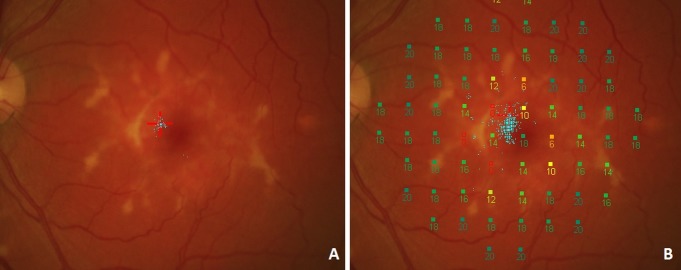
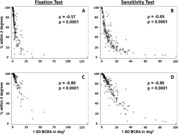
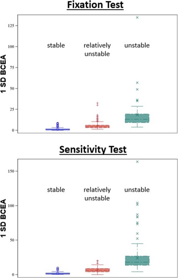
Fixation was tested during a separate fixation exam and also dynamically during a sensitivity exam, using fundus-tracking microperimetry (Nidek MP-1). Fixation data were analyzed using the bivariate contour ellipse area (BCEA), the 2/4 degree method, and the Fujii classification.

RESULTS

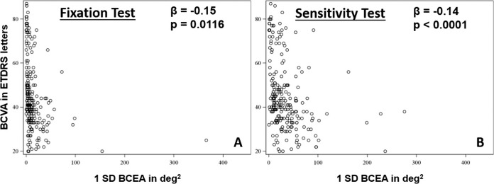
In a total of 235 patients, the mean BCEA was larger when measured during the sensitivity exam (418 eyes; 12.5 vs. 4.6 deg2 during the fixation task in 427 eyes). Correlations between the two tests were generally weak. Fixation stability during the sensitivity test was significantly correlated with visual acuity. Comparing the BCEA values and the corresponding Fujii categories for these eyes revealed ranges of overlap where an eye with one defined BCEA value can fall into each of the three Fujii categories.

CONCLUSIONS

Patients may have limited ability to fixate over defined time periods, which leads to significant differences between shorter and longer measurements of fixation stability. The most appropriate way to use this functional biomarker appears to be using continuous metrics for fixation stability, such as the BCEA, during a macular sensitivity test.

摘要

目的

在多中心前瞻性ProgStar研究中,比较不同指标和采集模式下的注视稳定性,将其作为一大群ABCA4相关斯塔加特病患者的一种新的视觉功能生物标志物。

方法

使用眼底跟踪微视野计(尼德克MP-1)在单独的注视检查期间以及在敏感度检查期间动态测试注视。使用双变量等高椭圆面积(BCEA)、2/4度法和藤井分类法分析注视数据。

结果

在总共235名患者中,在敏感度检查期间测量时,平均BCEA更大(418只眼;在427只眼中,注视任务期间为12.5度²,而在敏感度检查期间为4.6度²)。两项测试之间的相关性通常较弱。敏感度测试期间的注视稳定性与视力显著相关。比较这些眼睛的BCEA值和相应的藤井类别,发现存在重叠范围,即具有一个定义的BCEA值的眼睛可以落入三个藤井类别中的每一个。

结论

患者在规定时间段内的注视能力可能有限,这导致了注视稳定性的短期和长期测量之间存在显著差异。使用这种功能生物标志物的最合适方法似乎是在黄斑敏感度测试期间使用连续的注视稳定性指标,如BCEA。

https://cdn.ncbi.nlm.nih.gov/pmc/blobs/aa07/6733530/86c80ad089b2/i1552-5783-58-6-BIO268-f04.jpg
https://cdn.ncbi.nlm.nih.gov/pmc/blobs/aa07/6733530/92e5c0f8f1d2/i1552-5783-58-6-BIO268-f01.jpg
https://cdn.ncbi.nlm.nih.gov/pmc/blobs/aa07/6733530/57c9125ec89b/i1552-5783-58-6-BIO268-f02.jpg
https://cdn.ncbi.nlm.nih.gov/pmc/blobs/aa07/6733530/cc92d782b115/i1552-5783-58-6-BIO268-f03.jpg
https://cdn.ncbi.nlm.nih.gov/pmc/blobs/aa07/6733530/86c80ad089b2/i1552-5783-58-6-BIO268-f04.jpg

相似文献

1
Metrics and Acquisition Modes for Fixation Stability as a Visual Function Biomarker.作为视觉功能生物标志物的固定稳定性的指标和采集模式。
Invest Ophthalmol Vis Sci. 2017 May 1;58(6):BIO268-BIO276. doi: 10.1167/iovs.17-21710.
2
Longitudinal Changes of Fixation Location and Stability Within 12 Months in Stargardt Disease: ProgStar Report No. 12.Stargardt 病患者 12 个月内固视位置和稳定性的纵向变化:ProgStar 报告第 12 号。
Am J Ophthalmol. 2018 Sep;193:54-61. doi: 10.1016/j.ajo.2018.06.003. Epub 2018 Jun 8.
3
Macular Sensitivity Measured With Microperimetry in Stargardt Disease in the Progression of Atrophy Secondary to Stargardt Disease (ProgStar) Study: Report No. 7.在斯塔加特病萎缩进展期斯塔加特病(ProgStar)研究中使用微视野计测量黄斑敏感性:第7号报告。
JAMA Ophthalmol. 2017 Jul 1;135(7):696-703. doi: 10.1001/jamaophthalmol.2017.1162.
4
Factors Influencing Fixation Stability Area: A Comparison of Two Methods of Recording.影响固定稳定区域的因素:两种记录方法的比较
Optom Vis Sci. 2018 Apr;95(4):384-390. doi: 10.1097/OPX.0000000000001201.
5
The Effect of Attention on Fixation Stability During Dynamic Fixation Testing in Stargardt Disease.注意对斯塔加特病患者动态固视测试中固视稳定性的影响。
Am J Ophthalmol. 2020 Sep;217:305-316. doi: 10.1016/j.ajo.2020.05.002. Epub 2020 May 11.
6
Static and dynamic retinal fixation stability in microperimetry.静态和动态视网膜固视稳定性的微视野检查。
Can J Ophthalmol. 2013 Oct;48(5):375-80. doi: 10.1016/j.jcjo.2013.05.021.
7
Fixation stability measurements in patients with neovascular age-related macular degeneration treated with ranibizumab.接受雷珠单抗治疗的新生血管性年龄相关性黄斑变性患者的固定稳定性测量。
Can J Ophthalmol. 2013 Oct;48(5):394-9. doi: 10.1016/j.jcjo.2013.04.006.
8
Evaluation of fixation stability using different targets with the MP1 microperimeter.使用MP1微视野计结合不同靶点评估固视稳定性。
Int Ophthalmol. 2015 Feb;35(1):11-7. doi: 10.1007/s10792-014-9924-x. Epub 2014 Apr 11.
9
Nidek MP1 is able to detect subtle decline in function in inherited and age-related atrophic macular disease with stable visual acuity.尼德克 MP1 能够检测到遗传性和年龄相关性萎缩性黄斑病变中功能的细微下降,而这些病变的视力是稳定的。
Retina. 2011 Feb;31(2):371-9. doi: 10.1097/IAE.0b013e3181e46af3.
10
The Natural History of the Progression of Atrophy Secondary to Stargardt Disease (ProgStar) Studies: Design and Baseline Characteristics: ProgStar Report No. 1.《继发于斯塔加特病的萎缩自然史(ProgStar)研究:设计与基线特征》:ProgStar 报告第 1 号。
Ophthalmology. 2016 Apr;123(4):817-28. doi: 10.1016/j.ophtha.2015.12.009. Epub 2016 Jan 16.

引用本文的文献

1
The natural preferred retinal locus in patients with macular disease.黄斑疾病患者的自然视网膜偏好位点。
Sci Rep. 2025 Jul 14;15(1):25348. doi: 10.1038/s41598-025-88421-6.
2
The vertical position of visual information conditions spatial memory performance in healthy aging.视觉信息的垂直位置影响健康老年人的空间记忆表现。
Commun Psychol. 2023 Jul 25;1(1):2. doi: 10.1038/s44271-023-00002-3.
3
Eye Tracking in Optometry: A Systematic Review.验光中的眼动追踪:一项系统综述。

本文引用的文献

1
Progression of Visual Acuity and Fundus Autofluorescence in Recent-Onset Stargardt Disease: ProgStar Study Report #4.新近发病的Stargardt病患者的视力和眼底自发荧光进展:ProgStar研究报告#4
Ophthalmol Retina. 2017 Nov-Dec;1(6):514-523. doi: 10.1016/j.oret.2017.02.008. Epub 2017 Apr 28.
2
Fixation Location and Stability Using the MP-1 Microperimeter in Stargardt Disease: ProgStar Report No. 3.使用MP-1微视野计在Stargardt病中的固视位置与稳定性:ProgStar报告第3号
Ophthalmol Retina. 2017 Jan-Feb;1(1):68-76. doi: 10.1016/j.oret.2016.08.009. Epub 2016 Oct 31.
3
7-Hexagon Multifocal Electroretinography for an Objective Functional Assessment of the Macula in 14 Seconds.
J Eye Mov Res. 2023 Aug 16;16(3). doi: 10.16910/jemr.16.3.3. eCollection 2023.
4
Alterations in Fixation Indices in Primary Open-Angle Glaucoma by Microperimetry.微视野检查法检测原发性开角型青光眼注视指数的变化
J Clin Med. 2022 Apr 23;11(9):2368. doi: 10.3390/jcm11092368.
5
The role of multimodal imaging and vision function testing in -related retinopathies and their relevance to future therapeutic interventions.多模态成像和视觉功能测试在相关视网膜病变中的作用及其与未来治疗干预的相关性。
Ther Adv Ophthalmol. 2021 Dec 19;13:25158414211056384. doi: 10.1177/25158414211056384. eCollection 2021 Jan-Dec.
6
Clinical Perspectives and Trends: Microperimetry as a Trial Endpoint in Retinal Disease.临床观点与趋势:微视野计在视网膜疾病中的试验终点应用。
Ophthalmologica. 2021;244(5):418-450. doi: 10.1159/000515148. Epub 2021 Feb 10.
7
A Workshop on Measuring the Progression of Atrophy Secondary to Stargardt Disease in the ProgStar Studies: Findings and Lessons Learned.ProgStar研究中测量Stargardt病继发萎缩进展的研讨会:研究结果与经验教训
Transl Vis Sci Technol. 2019 Apr 12;8(2):16. doi: 10.1167/tvst.8.2.16. eCollection 2019 Apr.
8
Microperimetry for geographic atrophy secondary to age-related macular degeneration.年龄相关性黄斑变性继发地图样萎缩的微视野检查。
Surv Ophthalmol. 2019 May-Jun;64(3):353-364. doi: 10.1016/j.survophthal.2019.01.014. Epub 2019 Jan 28.
9
Inherited Retinal Degenerations: Current Landscape and Knowledge Gaps.遗传性视网膜变性:现状与知识空白
Transl Vis Sci Technol. 2018 Jul 18;7(4):6. doi: 10.1167/tvst.7.4.6. eCollection 2018 Jul.
10
Scotopic Microperimetric Assessment of Rod Function in Stargardt Disease (SMART) Study: Design and Baseline Characteristics (Report No. 1).斯塔加特病视杆细胞功能的暗视微视野评估(SMART)研究:设计与基线特征(报告第1号)
Ophthalmic Res. 2019;61(1):36-43. doi: 10.1159/000488711. Epub 2018 Jun 25.
7 六边形多焦视网膜电图在 14 秒内对黄斑进行客观功能评估
Ophthalmic Res. 2017;58(2):117-124. doi: 10.1159/000475996. Epub 2017 Jun 23.
4
Macular Sensitivity Measured With Microperimetry in Stargardt Disease in the Progression of Atrophy Secondary to Stargardt Disease (ProgStar) Study: Report No. 7.在斯塔加特病萎缩进展期斯塔加特病(ProgStar)研究中使用微视野计测量黄斑敏感性:第7号报告。
JAMA Ophthalmol. 2017 Jul 1;135(7):696-703. doi: 10.1001/jamaophthalmol.2017.1162.
5
Maximum pain on visual analog scales in spinal disorders.脊柱疾病视觉模拟量表上的最大疼痛程度。
Spine J. 2017 Aug;17(8):1061-1065. doi: 10.1016/j.spinee.2016.11.017. Epub 2016 Dec 6.
6
Emerging therapies for inherited retinal degeneration.遗传性视网膜变性的新兴疗法。
Sci Transl Med. 2016 Dec 7;8(368):368rv6. doi: 10.1126/scitranslmed.aaf2838.
7
FUNDUS AUTOFLUORESCENCE IN A SUBCLINICAL CASE OF BEST DISEASE.贝斯特病亚临床病例中的眼底自发荧光
Retin Cases Brief Rep. 2017;11 Suppl 1:S159-S162. doi: 10.1097/ICB.0000000000000390.
8
Stargardt disease: clinical features, molecular genetics, animal models and therapeutic options.斯塔加特病:临床特征、分子遗传学、动物模型及治疗选择
Br J Ophthalmol. 2017 Jan;101(1):25-30. doi: 10.1136/bjophthalmol-2016-308823. Epub 2016 Aug 4.
9
Visual Acuity Loss and Associated Risk Factors in the Retrospective Progression of Stargardt Disease Study (ProgStar Report No. 2).《斯塔加特病回顾性进展研究(ProgStar 报告 2 号)中的视力丧失及相关危险因素》。
Ophthalmology. 2016 Sep;123(9):1887-97. doi: 10.1016/j.ophtha.2016.05.027. Epub 2016 Jul 2.
10
The Natural History of the Progression of Atrophy Secondary to Stargardt Disease (ProgStar) Studies: Design and Baseline Characteristics: ProgStar Report No. 1.《继发于斯塔加特病的萎缩自然史(ProgStar)研究:设计与基线特征》:ProgStar 报告第 1 号。
Ophthalmology. 2016 Apr;123(4):817-28. doi: 10.1016/j.ophtha.2015.12.009. Epub 2016 Jan 16.