• 文献检索
  • 文档翻译
  • 深度研究
  • 学术资讯
  • Suppr Zotero 插件Zotero 插件
  • 邀请有礼
  • 套餐&价格
  • 历史记录
应用&插件
Suppr Zotero 插件Zotero 插件浏览器插件Mac 客户端Windows 客户端微信小程序
定价
高级版会员购买积分包购买API积分包
服务
文献检索文档翻译深度研究API 文档MCP 服务
关于我们
关于 Suppr公司介绍联系我们用户协议隐私条款
关注我们

Suppr 超能文献

核心技术专利:CN118964589B侵权必究
粤ICP备2023148730 号-1Suppr @ 2026

文献检索

告别复杂PubMed语法,用中文像聊天一样搜索,搜遍4000万医学文献。AI智能推荐,让科研检索更轻松。

立即免费搜索

文件翻译

保留排版,准确专业,支持PDF/Word/PPT等文件格式,支持 12+语言互译。

免费翻译文档

深度研究

AI帮你快速写综述,25分钟生成高质量综述,智能提取关键信息,辅助科研写作。

立即免费体验

斯塔加特病视杆细胞功能的暗视微视野评估(SMART)研究:设计与基线特征(报告第1号)

Scotopic Microperimetric Assessment of Rod Function in Stargardt Disease (SMART) Study: Design and Baseline Characteristics (Report No. 1).

作者信息

Strauss Rupert W, Kong Xiangrong, Bittencourt Millena G, Ho Alexander, Jha Anamika, Schönbach Etienne M, Ahmed Mohamed I, Muñoz Beatriz, Ervin Ann-Margret, Michaelides Michel, Birch David G, Sahel José-Alain, Sunness Janet S, Zrenner Eberhart, Bagheri Saghar, Ip Michael, Sadda SriniVas, West Sheila, Scholl Hendrik P N

机构信息

Wilmer Eye Institute, Johns Hopkins University, Baltimore, Maryland, USA.

Moorfields Eye Hospital NHS Foundation Trust, and UCL Institute of Ophthalmology, University College London, London, United Kingdom.

出版信息

Ophthalmic Res. 2019;61(1):36-43. doi: 10.1159/000488711. Epub 2018 Jun 25.

DOI:10.1159/000488711
PMID:29940588
原文链接:https://pmc.ncbi.nlm.nih.gov/articles/PMC7175756/
Abstract

PURPOSE

To describe the study design and characteristics at first visit of participants in the longitudinal Scotopic Microperimetric Assessment of Rod Function in Stargardt Disease (SMART) study.

METHODS

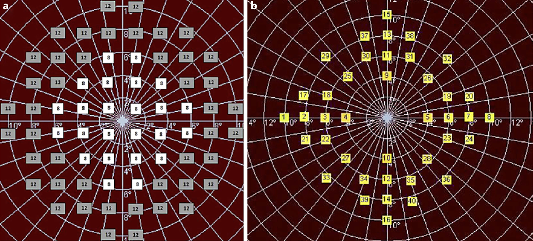
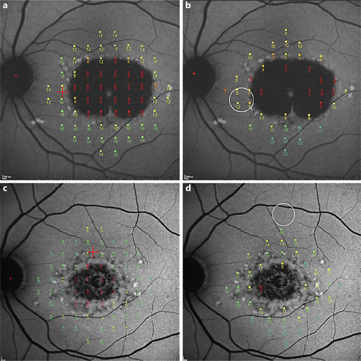
Scotopic microperimetry (sMP) was performed in one designated study eye in a subset of participants with molecularly proven ABCA4-associated Stargardt disease (STGD1) enrolled in a multicenter natural history study (ProgStar). Study visits were every 6 months over a period ranging from 6 to 24 months, and also included fundus autofluorescence (FAF).

RESULTS

SMART enrolled 118 participants (118 eyes). At the first visit of SMART, the mean sensitivity in mesopic microperimetry was 11.48 (±5.05; range 0.00-19.88) dB and in sMP 11.25 (±5.26; 0-19.25) dB. For FAF, all eyes had a lesion of decreased autofluorescence (mean lesion size 3.62 [±3.48; 0.10-21.46] mm2), and a total of 76 eyes (65.5%) had a lesion of definitely decreased autofluorescence with a mean lesion size of 3.46 (±3.60; 0.21-21.46) mm2.

CONCLUSIONS

Rod function is impaired in STGD1 and can be assessed by sMP. Testing rod function may serve as a potential outcome measure for future clinical treatment trials. This is evaluated in the SMART study.

摘要

目的

描述斯塔加特病视杆细胞功能纵向暗视微视野评估(SMART)研究中参与者首次就诊时的研究设计和特征。

方法

在一项多中心自然史研究(ProgStar)中,对一部分经分子学证实为ABCA4相关斯塔加特病(STGD1)的参与者的一只指定研究眼进行暗视微视野检查(sMP)。研究访视每6个月进行一次,为期6至24个月,还包括眼底自发荧光(FAF)检查。

结果

SMART研究招募了118名参与者(118只眼)。在SMART研究的首次就诊时,中视微视野检查的平均敏感度为11.48(±5.05;范围0.00 - 19.88)dB,sMP检查的平均敏感度为11.25(±5.26;0 - 19.25)dB。对于FAF检查,所有眼睛均有自发荧光降低的病变(平均病变面积3.62 [±3.48;0.10 - 21.46] mm²),共有76只眼(65.5%)有明确自发荧光降低的病变,平均病变面积为3.46(±3.60;0.21 - 21.46)mm²。

结论

STGD1患者的视杆细胞功能受损,可通过sMP进行评估。检测视杆细胞功能可能作为未来临床治疗试验的潜在结局指标。这在SMART研究中进行评估。

https://cdn.ncbi.nlm.nih.gov/pmc/blobs/58a2/7175756/9e530283cdec/nihms-1573836-f0002.jpg
https://cdn.ncbi.nlm.nih.gov/pmc/blobs/58a2/7175756/96fded6d367f/nihms-1573836-f0001.jpg
https://cdn.ncbi.nlm.nih.gov/pmc/blobs/58a2/7175756/9e530283cdec/nihms-1573836-f0002.jpg
https://cdn.ncbi.nlm.nih.gov/pmc/blobs/58a2/7175756/96fded6d367f/nihms-1573836-f0001.jpg
https://cdn.ncbi.nlm.nih.gov/pmc/blobs/58a2/7175756/9e530283cdec/nihms-1573836-f0002.jpg

相似文献

1
Scotopic Microperimetric Assessment of Rod Function in Stargardt Disease (SMART) Study: Design and Baseline Characteristics (Report No. 1).斯塔加特病视杆细胞功能的暗视微视野评估(SMART)研究:设计与基线特征(报告第1号)
Ophthalmic Res. 2019;61(1):36-43. doi: 10.1159/000488711. Epub 2018 Jun 25.
2
Macular Sensitivity Measured With Microperimetry in Stargardt Disease in the Progression of Atrophy Secondary to Stargardt Disease (ProgStar) Study: Report No. 7.在斯塔加特病萎缩进展期斯塔加特病(ProgStar)研究中使用微视野计测量黄斑敏感性:第7号报告。
JAMA Ophthalmol. 2017 Jul 1;135(7):696-703. doi: 10.1001/jamaophthalmol.2017.1162.
3
The Natural History of the Progression of Atrophy Secondary to Stargardt Disease (ProgStar) Studies: Design and Baseline Characteristics: ProgStar Report No. 1.《继发于斯塔加特病的萎缩自然史(ProgStar)研究:设计与基线特征》:ProgStar 报告第 1 号。
Ophthalmology. 2016 Apr;123(4):817-28. doi: 10.1016/j.ophtha.2015.12.009. Epub 2016 Jan 16.
4
Longitudinal Microperimetric Changes of Macular Sensitivity in Stargardt Disease After 12 Months: ProgStar Report No. 13.《Stargardt 病患者黄斑敏感性在 12 个月后的纵向微视野计变化:ProgStar 报告 No.13》。
JAMA Ophthalmol. 2020 Jul 1;138(7):772-779. doi: 10.1001/jamaophthalmol.2020.1735.
5
The Progression of the Stargardt Disease Type 4 (ProgStar-4) Study: Design and Baseline Characteristics (ProgStar-4 Report No. 1).斯塔加特病4型(ProgStar-4)研究进展:设计与基线特征(ProgStar-4报告第1号)
Ophthalmic Res. 2018;60(3):185-194. doi: 10.1159/000491791. Epub 2018 Aug 15.
6
Longitudinal Changes in Scotopic and Mesopic Macular Function as Assessed with Microperimetry in Patients With Stargardt Disease: SMART Study Report No. 2.《用微视野计评估斯塔加特病患者的暗点和中间视力黄斑功能的纵向变化:SMART 研究报告第 2 号》。
Am J Ophthalmol. 2022 Apr;236:32-44. doi: 10.1016/j.ajo.2021.10.014. Epub 2021 Oct 23.
7
Progression of Stargardt Disease as Determined by Fundus Autofluorescence in the Retrospective Progression of Stargardt Disease Study (ProgStar Report No. 9).在斯塔加特病回顾性进展研究(ProgStar报告第9号)中,通过眼底自发荧光确定的斯塔加特病进展情况。
JAMA Ophthalmol. 2017 Nov 1;135(11):1232-1241. doi: 10.1001/jamaophthalmol.2017.4152.
8
Retinal Sensitivity Using Microperimetry in Age-Related Macular Degeneration in an Amish Population.阿米什人群中年龄相关性黄斑变性患者使用微视野计测量视网膜敏感度
Ophthalmic Surg Lasers Imaging Retina. 2019 Sep 1;50(9):e236-e241. doi: 10.3928/23258160-20190905-15.
9
Morpho-functional correlation of fundus autofluorescence in Stargardt disease.Stargardt病中眼底自发荧光的形态功能相关性
Br J Ophthalmol. 2015 Oct;99(10):1354-9. doi: 10.1136/bjophthalmol-2014-306237. Epub 2015 Apr 2.
10
Visual Acuity Change Over 24 Months and Its Association With Foveal Phenotype and Genotype in Individuals With Stargardt Disease: ProgStar Study Report No. 10.24 个月期间的视力变化及其与斯塔加特病个体的中心凹表型和基因型的关系:ProgStar 研究报告第 10 号。
JAMA Ophthalmol. 2018 Aug 1;136(8):920-928. doi: 10.1001/jamaophthalmol.2018.2198.

引用本文的文献

1
Flavoprotein Fluorescence Imaging in Stargardt Disease: Linking Metabolic Stress to Structural Damage.斯塔加特病中的黄素蛋白荧光成像:将代谢应激与结构损伤联系起来。
Invest Ophthalmol Vis Sci. 2025 Aug 1;66(11):12. doi: 10.1167/iovs.66.11.12.
2
In-Depth Retinal Sensitivity Assessment With the MP3 Type S Microperimeter: A Methods Study.用 MP3 型 S 微视野计进行深入的视网膜灵敏度评估:一种方法研究。
Transl Vis Sci Technol. 2024 Apr 2;13(4):14. doi: 10.1167/tvst.13.4.14.
3
The Role of the Choroid in Stargardt Disease.脉络膜在斯塔加特病中的作用。

本文引用的文献

1
Fixation Location and Stability Using the MP-1 Microperimeter in Stargardt Disease: ProgStar Report No. 3.使用MP-1微视野计在Stargardt病中的固视位置与稳定性:ProgStar报告第3号
Ophthalmol Retina. 2017 Jan-Feb;1(1):68-76. doi: 10.1016/j.oret.2016.08.009. Epub 2016 Oct 31.
2
Metrics and Acquisition Modes for Fixation Stability as a Visual Function Biomarker.作为视觉功能生物标志物的固定稳定性的指标和采集模式。
Invest Ophthalmol Vis Sci. 2017 May 1;58(6):BIO268-BIO276. doi: 10.1167/iovs.17-21710.
3
Towards Treatment of Stargardt Disease: Workshop Organized and Sponsored by the Foundation Fighting Blindness.
Int J Mol Sci. 2022 Jul 9;23(14):7607. doi: 10.3390/ijms23147607.
4
Evaluation of Local Rod and Cone Function in Stargardt Disease.评估斯塔加特病中的局部杆状和锥状功能。
Invest Ophthalmol Vis Sci. 2022 Mar 2;63(3):6. doi: 10.1167/iovs.63.3.6.
5
The role of multimodal imaging and vision function testing in -related retinopathies and their relevance to future therapeutic interventions.多模态成像和视觉功能测试在相关视网膜病变中的作用及其与未来治疗干预的相关性。
Ther Adv Ophthalmol. 2021 Dec 19;13:25158414211056384. doi: 10.1177/25158414211056384. eCollection 2021 Jan-Dec.
6
Perifoveal Cone- and Rod-Mediated Temporal Contrast Sensitivities in Stargardt Disease/Fundus Flavimaculatus.Stargardt病/黄斑营养不良患者的中心凹周围视锥和视杆介导的时间对比敏感度
Invest Ophthalmol Vis Sci. 2021 Nov 1;62(14):24. doi: 10.1167/iovs.62.14.24.
7
Longitudinal Changes in Scotopic and Mesopic Macular Function as Assessed with Microperimetry in Patients With Stargardt Disease: SMART Study Report No. 2.《用微视野计评估斯塔加特病患者的暗点和中间视力黄斑功能的纵向变化:SMART 研究报告第 2 号》。
Am J Ophthalmol. 2022 Apr;236:32-44. doi: 10.1016/j.ajo.2021.10.014. Epub 2021 Oct 23.
8
Macular Rod Function in Retinitis Pigmentosa Measured With Scotopic Microperimetry.用暗视野微视野计测量色素性视网膜炎的黄斑杆功能。
Transl Vis Sci Technol. 2021 Sep 1;10(11):3. doi: 10.1167/tvst.10.11.3.
9
Clinical Perspectives and Trends: Microperimetry as a Trial Endpoint in Retinal Disease.临床观点与趋势:微视野计在视网膜疾病中的试验终点应用。
Ophthalmologica. 2021;244(5):418-450. doi: 10.1159/000515148. Epub 2021 Feb 10.
10
Randomised study evaluating the pharmacodynamics of emixustat hydrochloride in subjects with macular atrophy secondary to Stargardt disease.随机研究评估盐酸依美斯汀在继发于 Stargardt 病的黄斑萎缩患者中的药效动力学。
Br J Ophthalmol. 2022 Mar;106(3):403-408. doi: 10.1136/bjophthalmol-2020-317712. Epub 2020 Nov 19.
迈向斯塔加特病的治疗:由抗击失明基金会组织并赞助的研讨会
Transl Vis Sci Technol. 2017 Sep 14;6(5):6. doi: 10.1167/tvst.6.5.6. eCollection 2017 Sep.
4
Visual Acuity Change over 12 Months in the Prospective Progression of Atrophy Secondary to Stargardt Disease (ProgStar) Study: ProgStar Report Number 6.12 个月内 Stargardt 病相关萎缩的前瞻性进展(ProgStar)研究中视力的变化:ProgStar 报告第 6 号。
Ophthalmology. 2017 Nov;124(11):1640-1651. doi: 10.1016/j.ophtha.2017.04.026. Epub 2017 May 23.
5
Incidence of Atrophic Lesions in Stargardt Disease in the Progression of Atrophy Secondary to Stargardt Disease (ProgStar) Study: Report No. 5.斯塔加特病萎缩进展研究(ProgStar)中斯塔加特病萎缩性病变的发生率:第5号报告
JAMA Ophthalmol. 2017 Jul 1;135(7):687-695. doi: 10.1001/jamaophthalmol.2017.1121.
6
Macular Sensitivity Measured With Microperimetry in Stargardt Disease in the Progression of Atrophy Secondary to Stargardt Disease (ProgStar) Study: Report No. 7.在斯塔加特病萎缩进展期斯塔加特病(ProgStar)研究中使用微视野计测量黄斑敏感性:第7号报告。
JAMA Ophthalmol. 2017 Jul 1;135(7):696-703. doi: 10.1001/jamaophthalmol.2017.1162.
7
Emerging therapies for inherited retinal degeneration.遗传性视网膜变性的新兴疗法。
Sci Transl Med. 2016 Dec 7;8(368):368rv6. doi: 10.1126/scitranslmed.aaf2838.
8
Stargardt disease: clinical features, molecular genetics, animal models and therapeutic options.斯塔加特病:临床特征、分子遗传学、动物模型及治疗选择
Br J Ophthalmol. 2017 Jan;101(1):25-30. doi: 10.1136/bjophthalmol-2016-308823. Epub 2016 Aug 4.
9
Visual Acuity Loss and Associated Risk Factors in the Retrospective Progression of Stargardt Disease Study (ProgStar Report No. 2).《斯塔加特病回顾性进展研究(ProgStar 报告 2 号)中的视力丧失及相关危险因素》。
Ophthalmology. 2016 Sep;123(9):1887-97. doi: 10.1016/j.ophtha.2016.05.027. Epub 2016 Jul 2.
10
Comparison of Short-Wavelength Reduced-Illuminance and Conventional Autofluorescence Imaging in Stargardt Macular Dystrophy.短波长降低照度与传统自发荧光成像在斯塔加特黄斑营养不良中的比较
Am J Ophthalmol. 2016 Aug;168:269-278. doi: 10.1016/j.ajo.2016.06.003. Epub 2016 Jun 11.