Awad Eman A, Abou Samra Waleed A, Torky Magda A, El-Kannishy Amr M
Ophthalmology Center, Faculty of Medicine, Mansoura University, 24 Al-Gomhoria street, Mansoura, Egypt.
BMC Ophthalmol. 2017 Oct 6;17(1):186. doi: 10.1186/s12886-017-0584-2.
Keratoconus (KC) is usually a bilateral corneal ectatic disease. For significant asymmetric presentation (so called unilateral KC), the fellow eye has the mildest and earliest form of the disease, which is typically called forme fruste keratoconus. The aim of this study was to evaluate the sensitivity and specificity of parameters derived from a Scheimpflug imaging system (Pentacam) as well as the changes in the quality of mesopic vision in the apparently normal fellow eye (forme fruste) to detect the earliest and most sensitive parameters.
Patients with clinical keratoconus in one eye and forme fruste keratoconus in the fellow eye were compared to subjects with normal eyes. The patients were examined using a rotating Scheimpflug imaging system (Pentacam).The following parameters were evaluated: keratometry, minimum corneal thickness, pachymetry progression index (PPI), Ambrósio relational thickness (ART), posterior elevation, back difference elevation (BDE) and multimetric D index(D index). Receiver operating characteristic (ROC) curves were analyzed by evaluating the area under the curve (AUC) to detect the sensitivity and specificity of each parameter. Mesopic vision evaluations were performed by contrast sensitivity and glare tests for each group.
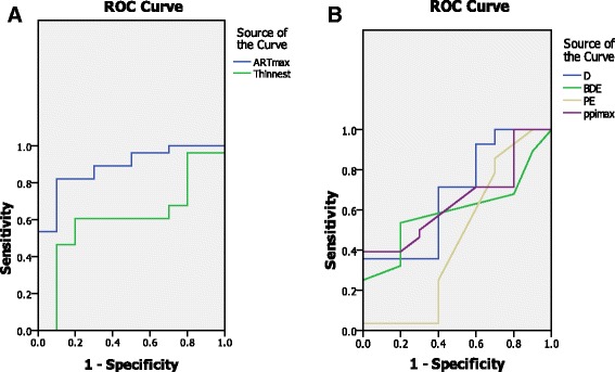
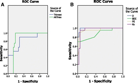
A total of 48 patients with clinical keratoconus in one eye and forme fruste keratoconus in the fellow eye and 72normal subjects were evaluated. In the clinical keratoconus eyes, the mean K, back difference elevation (BDE), pachymetric progression index maximum(PPI max), and multimetric D were significantly higher compared to the normal subjects, whereas the corneal pachymetry and Ambrósio relational thickness maximum (ART max) were significantly lower. In the forme fruste eyes, the ROC analysis showed that the AUC values of the mean K, thinnest pachymetry, ARTmax, BDE, D index, and PPI max were 0.82, 0.61, 0.88, 0. 67, and 0.64, respectively. The contrast sensitivity and glare tests were significantly affected in the forme fruste cases.
In forme fruste keratoconus eyes, the ART max is considered a highly sensitive objective parameter. Contrast sensitivity and glare is an important subjective test, which is affected in forme fruste patients.
圆锥角膜(KC)通常是一种双侧角膜扩张性疾病。对于显著不对称表现(即所谓的单侧KC),对侧眼患有该病最轻微和最早的形式,通常称为顿挫型圆锥角膜。本研究的目的是评估源自眼前节分析系统(Pentacam)的参数的敏感性和特异性,以及在看似正常的对侧眼(顿挫型)中,中间视觉质量的变化,以检测最早和最敏感的参数。
将一只眼睛患有临床圆锥角膜且对侧眼患有顿挫型圆锥角膜的患者与正常眼的受试者进行比较。使用旋转式眼前节分析系统(Pentacam)对患者进行检查。评估以下参数:角膜曲率测量、最小角膜厚度、角膜厚度进展指数(PPI)、安布罗西奥相关厚度(ART)、后表面高度、后表面高度差(BDE)和多指标D指数(D指数)。通过评估曲线下面积(AUC)分析受试者工作特征(ROC)曲线,以检测每个参数的敏感性和特异性。对每组进行中间视觉评估,包括对比敏感度和眩光测试。
共评估了48例一只眼睛患有临床圆锥角膜且对侧眼患有顿挫型圆锥角膜的患者和72名正常受试者。在临床圆锥角膜眼中,平均角膜曲率、后表面高度差(BDE)、角膜厚度进展指数最大值(PPI max)和多指标D指数均显著高于正常受试者,而角膜厚度和安布罗西奥相关厚度最大值(ART max)显著低于正常受试者。在顿挫型圆锥角膜眼中,ROC分析显示平均角膜曲率、最薄角膜厚度、ART max、BDE、D指数和PPI max的AUC值分别为0.82、0.61、0.88、0.67和0.64。在顿挫型圆锥角膜病例中,对比敏感度和眩光测试受到显著影响。
在顿挫型圆锥角膜眼中,ART max被认为是一个高度敏感的客观参数。对比敏感度和眩光测试是一项重要的主观测试,在顿挫型圆锥角膜患者中受到影响。