Vasudeva Viren S, Abd-El-Barr Muhammad, Pompeu Yuri A, Karhade Aditya, Groff Michael W, Lu Yi
Brigham and Women's Hospital, Harvard Medical School, Boston, MA, USA.
Global Spine J. 2017 Oct;7(7):648-656. doi: 10.1177/2192568217700100. Epub 2017 May 31.
Review and technical report.
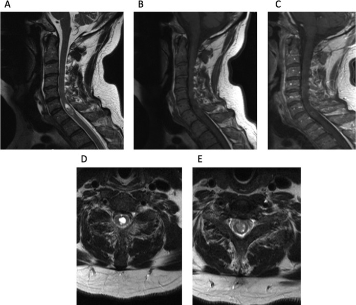
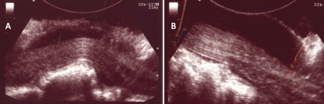
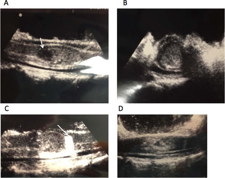
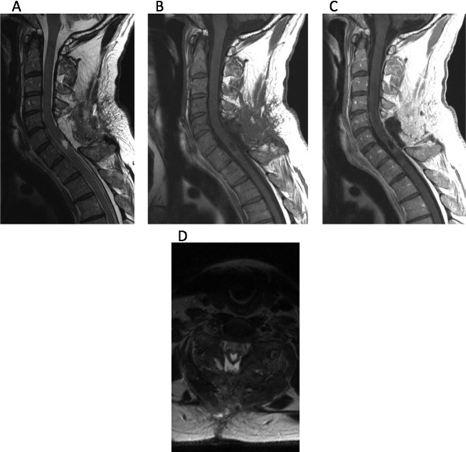
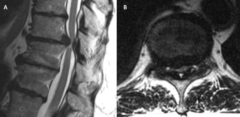
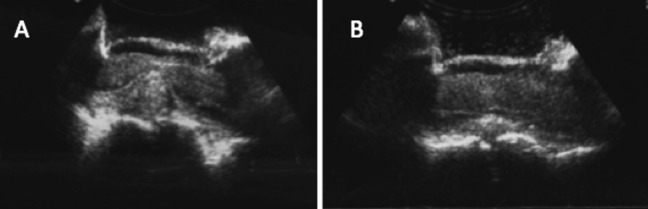
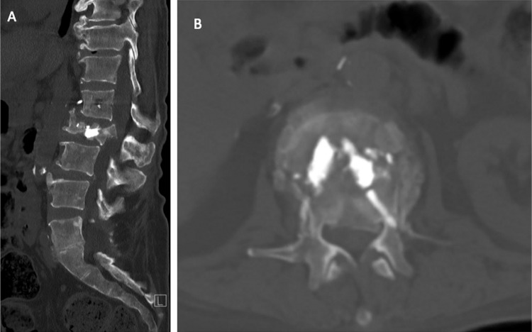
Intraoperative ultrasound has been used by spine surgeons since the early 1980s. Since that time, more advanced modes of intraoperative imaging and navigation have become widely available. Although the use of ultrasound during spine surgery has fallen out of favor, it remains the only true real-time imaging modality that allows surgeons to visualize soft tissue anatomy instantly and continuously while operating. It is our objective to demonstrate that for this reason, ultrasound is a useful adjunctive technique for spine surgeons, especially when approaching intradural lesions or when addressing pathology in the ventral spinal canal via a posterior approach.
Using PubMed, the existing literature regarding the use of intraoperative ultrasound during spinal surgery was evaluated. Also, surgical case logs were reviewed to identify spinal operations during which intraoperative ultrasound was used. Illustrative cases were selected and reviewed in detail.
This article provides a brief review of the history of intraoperative ultrasound in spine surgery and describes certain surgical scenarios during which this technique might be useful. Several illustrative cases are provided from our own experience.
Surgeons should consider the use of intraoperative ultrasound when approaching intradural lesions or when addressing pathology ventral to the thecal sac via a posterior approach.
综述与技术报告。
自20世纪80年代初以来,脊柱外科医生就开始使用术中超声。从那时起,更先进的术中成像和导航模式已广泛应用。尽管脊柱手术中超声的使用已不再流行,但它仍然是唯一真正的实时成像方式,使外科医生在手术过程中能够即时且持续地观察软组织解剖结构。我们的目的是证明,基于这个原因,超声对于脊柱外科医生是一种有用的辅助技术,特别是在处理硬膜内病变或通过后路处理脊髓腹侧椎管内病变时。
利用PubMed对脊柱手术中使用术中超声的现有文献进行评估。此外,回顾手术病例记录以确定使用术中超声的脊柱手术。选择并详细回顾了一些说明性病例。
本文简要回顾了脊柱手术中术中超声的历史,并描述了该技术可能有用的某些手术场景。提供了一些来自我们自身经验的说明性病例。
当处理硬膜内病变或通过后路处理硬膜囊腹侧病变时,外科医生应考虑使用术中超声。