Bednarik Petr, Moheet Amir A, Grohn Heidi, Kumar Anjali F, Eberly Lynn E, Seaquist Elizabeth R, Mangia Silvia
Department of Radiology, Center for Magnetic Resonance Research, University of MinnesotaMinneapolis, MN, United States.
Department of Medicine, University of MinnesotaMinneapolis, MN, United States.
Front Neurosci. 2017 Sep 25;11:529. doi: 10.3389/fnins.2017.00529. eCollection 2017.
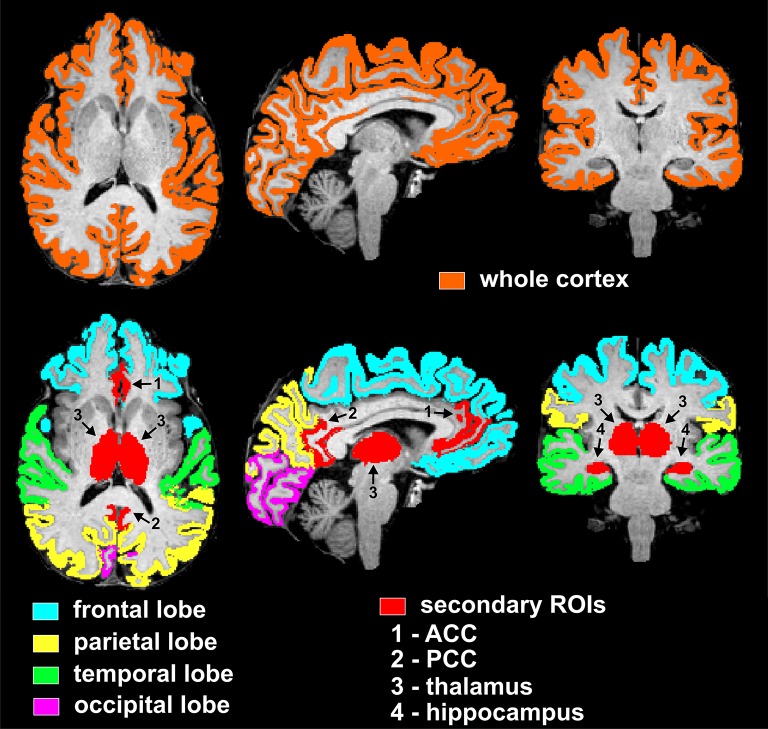
In this study, we retrospectively analyzed the anatomical MRI data acquired from 52 subjects with type 1 diabetes (26M/26F, 36 ± 11 years old, A1C = 7.2 ± 0.9%) and 50 age, sex and BMI frequency-matched non-diabetic controls (25M/25F, 36 ± 14 years old). The T1D group was further sub-divided based on whether subjects had normal, impaired, or indeterminate awareness of hypoglycemia ( = 31, 20, and 1, respectively). Our goals were to test whether the gray matter (GM) volumes of selected brain regions were associated with diabetes status as well as with the status of hypoglycemia awareness. T1D subjects were found to have slightly smaller volume of the whole cortex as compared to controls (-2.7%, = 0.016), with the most affected brain region being the frontal lobe (-3.6%, = 0.024). Similar differences of even larger magnitude were observed among the T1D subjects based on their hypoglycemia awareness status. Indeed, compared to the patients with normal awareness of hypoglycemia, patients with impaired awareness had smaller volume of the whole cortex (-7.9%, = 0.0009), and in particular of the frontal lobe (-9.1%, = 0.006), parietal lobe (-8.0%, = 0.015) and temporal lobe (-8.2%, = 0.009). Such differences were very similar to those observed between patients with impaired awareness and controls (-7.6%, = 0.0002 in whole cortex, -9.1%, = 0.0003 in frontal lobe, -7.8%, = 0.002 in parietal lobe, and -6.4%, = 0.019 in temporal lobe). On the other hand, patients with normal awareness did not present significant volume differences compared to controls. No group-differences were observed in the occipital lobe or in the anterior cingulate, posterior cingulate, hippocampus, and thalamus. We conclude that diabetes status is associated with a small but statistically significant reduction of the whole cortex volume, mainly in the frontal lobe. The most prominent structural effects occurred in patients with impaired awareness of hypoglycemia (IAH) as compared to those with normal awareness, perhaps due to the long-term exposure to recurrent episodes of hypoglycemia. Future studies aimed at quantifying relationships of structural outcomes with functional outcomes, with cognitive performance, as well as with parameters describing glucose variability and severity of hypoglycemia episodes, will be necessary to further understand the impact of T1D on the brain.
在本研究中,我们回顾性分析了52例1型糖尿病患者(26例男性/26例女性,年龄36±11岁,糖化血红蛋白[A1C]=7.2±0.9%)以及50例年龄、性别和体重指数(BMI)频率匹配的非糖尿病对照者(25例男性/25例女性,年龄36±14岁)的解剖学磁共振成像(MRI)数据。1型糖尿病组根据受试者低血糖意识正常、受损或不确定进一步细分(分别为31例、20例和1例)。我们的目标是测试选定脑区的灰质(GM)体积是否与糖尿病状态以及低血糖意识状态相关。结果发现,与对照组相比,1型糖尿病受试者的整个皮质体积略小(-2.7%,P=0.016),受影响最严重的脑区是额叶(-3.6%,P=0.024)。根据低血糖意识状态,在1型糖尿病受试者中观察到了更大幅度的类似差异。事实上,与低血糖意识正常的患者相比,意识受损的患者整个皮质体积更小(-7.9%,P=0.0009),尤其是额叶(-9.1%,P=0.006)、顶叶(-8.0%,P=0.015)和颞叶(-8.2%,P=0.009)。这些差异与意识受损患者和对照组之间观察到的差异非常相似(整个皮质为-7.6%,P=0.0002;额叶为-9.1%,P=0.0003;顶叶为-7.8%,P=0.002;颞叶为-6.4%,P=0.019)。另一方面,低血糖意识正常的患者与对照组相比,未出现显著的体积差异。在枕叶或前扣带回、后扣带回、海马体和丘脑未观察到组间差异。我们得出结论,糖尿病状态与整个皮质体积的微小但具有统计学意义的减少相关,主要在额叶。与意识正常的患者相比,低血糖意识受损(IAH)患者出现的结构影响最为显著,这可能是由于长期暴露于反复发生的低血糖发作所致。未来有必要开展旨在量化结构结果与功能结果、认知表现以及描述血糖变异性和低血糖发作严重程度的参数之间关系的研究,以进一步了解1型糖尿病对大脑的影响。