Itaya Sakiko, Ueda Yasuhiro, Kobayashi Zen, Tomimitsu Hiroyuki, Kobayashi Daisuke, Shintani Shuzo
Department of Neurology, JA Toride Medical Center, Japan.
Department of Neurosurgery, JA Toride Medical Center, Japan.
Intern Med. 2017 Dec 15;56(24):3353-3355. doi: 10.2169/internalmedicine.8832-17. Epub 2017 Oct 11.
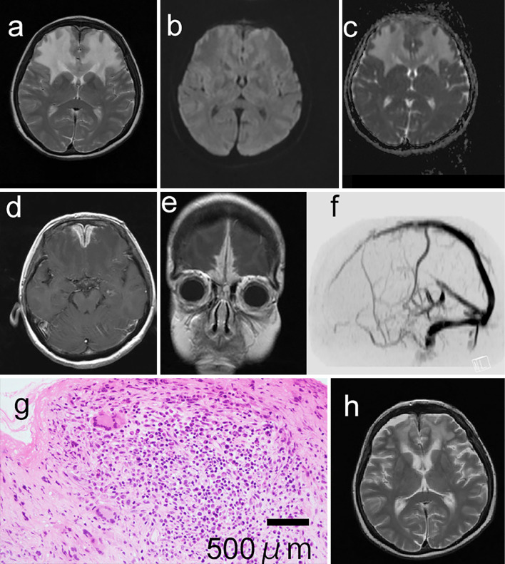
A 61-year-old woman presented with a 1-month history of decreased activities of daily living. Magnetic resonance imaging revealed abnormal intensities of the bilateral frontal lobes and enhancement of the thickened dura matter. A biopsy of the dura mater revealed multinucleated giant cells. She had sinusitis and hematuria; she was diagnosed with granulomatosis with polyangiitis. Hypertrophic pachymeningitis (HPM) was considered to have interrupted the venous flow and caused vasogenic edema. Bilateral frontal lobe edema resulting from HPM due to granulomatosis with polyangiitis has not been reported. A biopsy and examination for other organ complications were useful for the diagnosis and treatment of our patient.
一名61岁女性因日常生活活动能力下降1个月前来就诊。磁共振成像显示双侧额叶信号异常,硬脑膜增厚并强化。硬脑膜活检发现多核巨细胞。她患有鼻窦炎和血尿,被诊断为肉芽肿性多血管炎。肥厚性硬脑膜炎(HPM)被认为中断了静脉血流并导致血管源性水肿。肉芽肿性多血管炎所致HPM引起的双侧额叶水肿尚未见报道。对我们的患者进行活检及其他器官并发症检查对诊断和治疗很有用。