Ologun Gabriel O, Patel Zinal M, Rana Navpreet K, Trecartin Andrew, Shen Alice, Trostle Douglas, Bertsch David
General Surgery, Guthrie Clinic/Robert Packer Hospital.
Medicine, Winthrop University Hospital.
Cureus. 2017 Aug 9;9(8):e1552. doi: 10.7759/cureus.1552.
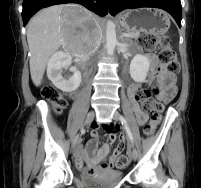
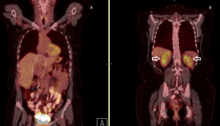
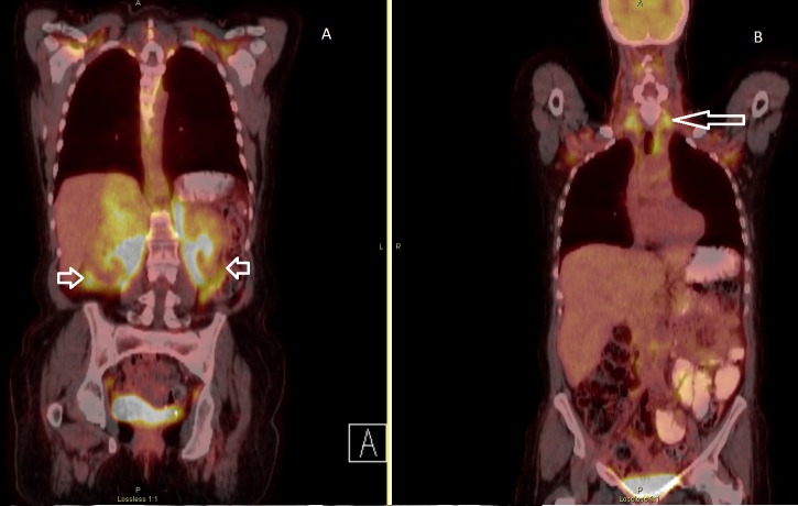
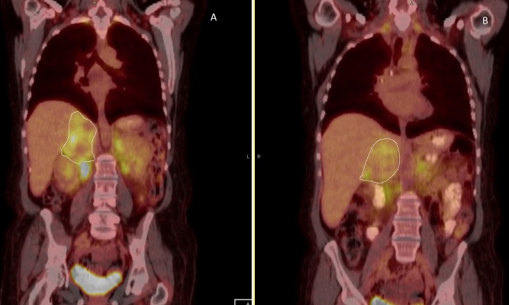
Pheochromocytomas are rare tumors derived from chromaffin cells located in the adrenal and extra adrenal tissues. Pheochromocytomas are diagnosed biochemically and localized using different imaging modalities. The definitive management is surgical resection. Brown adipose tissues are normally present during fetal development, with regression over time. Brown adipose tissues are thermogenic and usually located in the neck, mediastinum, and retroperitoneum. Here, we report a case of a unilateral pheochromocytoma surrounded by brown fat. The abnormal stimulation of brown fat noted on positive emission tomography scan (PET) resolved after the pheochromocytoma was resected.
嗜铬细胞瘤是起源于肾上腺和肾上腺外组织中嗜铬细胞的罕见肿瘤。嗜铬细胞瘤通过生化方法诊断,并使用不同的成像方式进行定位。明确的治疗方法是手术切除。棕色脂肪组织通常在胎儿发育期间存在,随着时间推移会退化。棕色脂肪组织具有产热功能,通常位于颈部、纵隔和腹膜后。在此,我们报告一例单侧嗜铬细胞瘤被棕色脂肪包绕的病例。嗜铬细胞瘤切除后,正电子发射断层扫描(PET)上显示的棕色脂肪异常刺激消失。