Abassi Amir Jalal, Mohamadnia Abdolreza, Parhiz Seyed Alireza, Azizi Moghadam Nahid, Bahrami Naghmeh
Craniomaxillofacial Research Center, Tehran University of Medical Sciences, Tehran, Iran.
Virology Research Center, National Research Institute of Tuberculosis and Lung Diseases (NRITLD), Shahid Beheshti University of Medical Sciences, Tehran, Iran.
J Dent (Shiraz). 2017 Sep;18(3):219-226.
Evidence shows thiabendazole has the potential to inhibit angiogenesis in melanoma and fibrosarcoma; however, its effect on oral squamous cell carcinoma has not been previously studied.
This study sought to assess the cytotoxic effects of thiabendazole on HN5 head and neck squamous carcinoma cell line.
HN5 cell lines were exposed to different concentrations of thiabendazole (prepared from 99% pure powder) for 24, 48 and 72 hours. Cell viability was assessed by the methyl thiazol tetrazolium assay, and IC50 of thiabendazole was calculated. Cells were also exposed to different concentrations of thiabendazole for 48 hours to determine its effect on expression and transcription of vascular endothelial growth factor gene. Expression of vascular endothelial growth factor mRNA was assessed by real-time polymerase chain reaction. The vascular endothelial growth factor release was assessed by the enzyme-linked immunosorbent assay test.
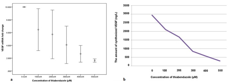
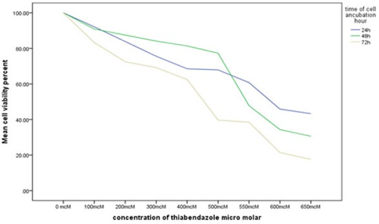
In all concentrations of thiabendazole except for 200 and 550μM, cell viability was significantly different at different time points (< 0.05). At 48 and 72 hours, cell viability at all concentrations of thiabendazole (100-650μM) significantly decreased compared to the control group (zero concentration). In addition, cell viability significantly decreased with an increase in thiabendazole concentration. At 48 hours, expression of vascular endothelial growth factor mRNA was significantly lower in presence of 500μM thiabendazole compared to the control group (< 0.001) and release of vascular endothelial growth factor was inhibited in a dose-dependent manner.
Thiabendazole inhibited the proliferation of HN5 cells in a dose-dependent and time-dependent manner. It also inhibited the expression of vascular endothelial growth factor gene.
有证据表明噻苯达唑有可能抑制黑色素瘤和纤维肉瘤中的血管生成;然而,其对口腔鳞状细胞癌的影响此前尚未得到研究。
本研究旨在评估噻苯达唑对HN5头颈部鳞状癌细胞系的细胞毒性作用。
将HN5细胞系暴露于不同浓度的噻苯达唑(由99%的纯粉末制备)中24、48和72小时。通过甲基噻唑四氮唑法评估细胞活力,并计算噻苯达唑的半数抑制浓度(IC50)。细胞还被暴露于不同浓度的噻苯达唑中48小时,以确定其对血管内皮生长因子基因表达和转录的影响。通过实时聚合酶链反应评估血管内皮生长因子mRNA的表达。通过酶联免疫吸附测定试验评估血管内皮生长因子的释放。
除200和550μM外,在所有浓度的噻苯达唑中,不同时间点的细胞活力均有显著差异(<0.05)。在48和72小时时,与对照组(零浓度)相比,所有浓度(100 - 650μM)的噻苯达唑处理下细胞活力均显著降低。此外,细胞活力随噻苯达唑浓度的增加而显著降低。在48小时时,与对照组相比,500μM噻苯达唑存在时血管内皮生长因子mRNA的表达显著降低(<0.001),且血管内皮生长因子的释放受到剂量依赖性抑制。
噻苯达唑以剂量和时间依赖性方式抑制HN5细胞的增殖。它还抑制血管内皮生长因子基因的表达。