Department of Cardiology, Medical University Hospital Heidelberg, INF 410, D-69120, Heidelberg, Germany.
DZHK (German Centre for Cardiovascular Research), partner site Heidelberg/Mannheim, University of Heidelberg, INF 410, D-69120, Heidelberg, Germany.
Stem Cell Res Ther. 2017 Oct 16;8(1):229. doi: 10.1186/s13287-017-0681-4.
Human induced pluripotent stem cells (hiPSC) harbor the potential to differentiate into diverse cardiac cell types. Previous experimental efforts were primarily directed at the generation of hiPSC-derived cells with ventricular cardiomyocyte characteristics. Aiming at a straightforward approach for pacemaker cell modeling and replacement, we sought to selectively differentiate cells with nodal-type properties.
hiPSC were differentiated into spontaneously beating clusters by co-culturing with visceral endoderm-like cells in a serum-free medium. Subsequent culturing in a specified fetal bovine serum (FBS)-enriched cell medium produced a pacemaker-type phenotype that was studied in detail using quantitative real-time polymerase chain reaction (qRT-PCR), immunocytochemistry, and patch-clamp electrophysiology. Further investigations comprised pharmacological stimulations and co-culturing with neonatal cardiomyocytes.
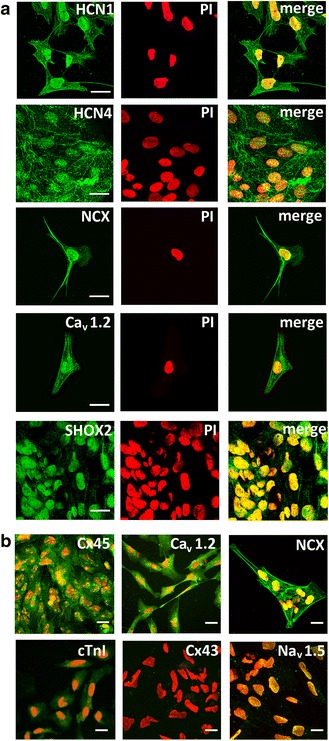
hiPSC co-cultured in a serum-free medium with the visceral endoderm-like cell line END-2 produced spontaneously beating clusters after 10-12 days of culture. The pacemaker-specific genes HCN4, TBX3, and TBX18 were abundantly expressed at this early developmental stage, while levels of sarcomeric gene products remained low. We observed that working-type cardiomyogenic differentiation can be suppressed by transfer of early clusters into a FBS-enriched cell medium immediately after beating onset. After 6 weeks under these conditions, sinoatrial node (SAN) hallmark genes remained at high levels, while working-type myocardial transcripts (NKX2.5, TBX5) were low. Clusters were characterized by regular activity and robust beating rates (70-90 beats/min) and were triggered by spontaneous Ca transients recapitulating calcium clock properties of genuine pacemaker cells. They were responsive to adrenergic/cholinergic stimulation and able to pace neonatal rat ventricular myocytes in co-culture experiments. Action potential (AP) measurements of cells individualized from clusters exhibited nodal-type (63.4%) and atrial-type (36.6%) AP morphologies, while ventricular AP configurations were not observed.
We provide a novel culture media-based, transgene-free approach for targeted generation of hiPSC-derived pacemaker-type cells that grow in clusters and offer the potential for disease modeling, drug testing, and individualized cell-based replacement therapy of the SAN.
人类诱导多能干细胞(hiPSC)具有分化为多种心脏细胞类型的潜力。先前的实验研究主要集中在产生具有心室肌细胞特征的 hiPSC 衍生细胞上。为了直接模拟和替代起搏细胞,我们试图选择性地分化具有结型特性的细胞。
通过与类内脏内胚层细胞共培养,在无血清培养基中使 hiPSC 分化为自发搏动的细胞簇。随后在特定的胎牛血清(FBS)丰富的细胞培养基中培养,产生起搏细胞类型的表型,并用实时定量聚合酶链反应(qRT-PCR)、免疫细胞化学和膜片钳电生理学进行详细研究。进一步的研究包括药物刺激和与新生心肌细胞共培养。
hiPSC 在无血清培养基中与类内脏内胚层细胞系 END-2 共培养 10-12 天后产生自发搏动的细胞簇。在这个早期发育阶段,起搏特异性基因 HCN4、TBX3 和 TBX18 大量表达,而肌节基因产物的水平仍然较低。我们观察到,通过在搏动开始后立即将早期细胞簇转移到 FBS 丰富的细胞培养基中,可以抑制工作型心肌生成分化。在这些条件下培养 6 周后,窦房结(SAN)标志性基因仍保持高水平,而工作型心肌转录物(NKX2.5、TBX5)水平较低。细胞簇表现出规则的活动和强劲的搏动率(70-90 次/分钟),并通过自发钙瞬变触发,再现了真正起搏细胞的钙钟特性。它们对肾上腺素能/胆碱能刺激有反应,并能够在共培养实验中起搏新生大鼠心室肌细胞。从细胞簇中分离的单个细胞的动作电位(AP)测量表现出结型(63.4%)和心房型(36.6%)AP 形态,而未观察到心室 AP 构型。
我们提供了一种新型的基于培养介质、无转基因的方法,用于靶向生成 hiPSC 衍生的起搏细胞类型,这些细胞在簇中生长,并为疾病建模、药物测试和 SAN 的个体化细胞替代治疗提供了潜力。