Genomics, Proteomics and Biomedical Functions, Department of Life and Medical Systems, Faculty of Life and Medical Sciences, Doshisha University, Kyoto, Japan.
Neuropathology, Department of Life and Medical Systems, Faculty of Life and Medical Sciences, Doshisha University, Kyoto, Japan.
Acta Neuropathol Commun. 2017 Oct 16;5(1):73. doi: 10.1186/s40478-017-0477-x.
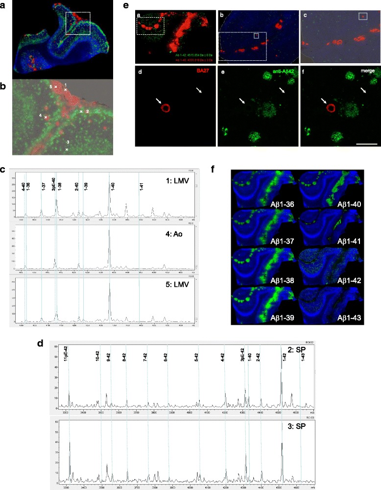
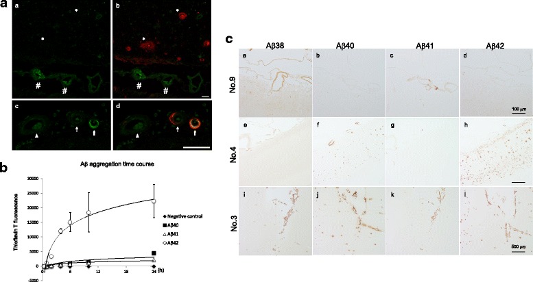
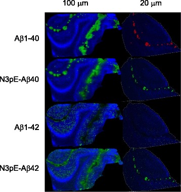
Amyloid β (Aβ) deposition in the brain is an early and invariable feature of Alzheimer's disease (AD). The Aβ peptides are composed of about 40 amino acids and are generated from amyloid precursor proteins (APP), by β- and γ-secretases. The distribution of individual Aβ peptides in the brains of aged people, and those suffering from AD and cerebral amyloid angiopathy (CAA), is not fully characterized. We employed the matrix-assisted laser desorption/ionization-imaging mass spectrometry (MALDI-IMS) to illustrate the spatial distribution of a broad range of Aβ species in human autopsied brains. With technical advancements such as formic acid pretreatment of frozen autopsied brain samples, we have: i) demonstrated that Aβ1-42 and Aβ1-43 were selectively deposited in senile plaques while full-length Aβ peptides such as Aβ1-36, 1-37, 1-38, 1-39, 1-40, and Aβ1-41 were deposited in leptomeningeal blood vessels. ii) Visualized distinct depositions of N-terminal truncated Aβ40 and Aβ42, including pyroglutamate modified at Glu-3 (N3pE), only with IMS for the first time. iii) Demonstrated that one single amino acid alteration at the C-terminus between Aβ1-42 and Aβ1-41 results in profound changes in their distribution pattern. In vitro, this can be attributed to the difference in the self-aggregation ability amongst Aβ1-40, Aβ1-41, and Aβ1-42. These observations were further confirmed with immunohistochemistry (IHC), using the newly developed anti-Aβ1-41 antibody. Here, distinct depositions of truncated and/or modified C- and N-terminal fragments of Aβs in AD and CAA brains with MALDI-IMS were visualized in a spacio-temporal specific manner. Specifically, Aβ1-41 was detected both with MALDI-IMS and IHC suggesting that a single amino acid alteration at the C-terminus of Aβ results in drastic distribution changes. These results suggest that MALDI-IMS could be used as a standard approach in combination with clinical, genetic, and pathological observations in understanding the pathology of AD and CAA.
脑内淀粉样蛋白 β(Aβ)沉积是阿尔茨海默病(AD)的早期和不变特征。Aβ 肽由约 40 个氨基酸组成,由淀粉样前体蛋白(APP)通过β-和γ-分泌酶产生。在老年人、AD 和脑淀粉样血管病(CAA)患者的大脑中,个体 Aβ 肽的分布尚未完全确定。我们采用基质辅助激光解吸/电离成像质谱(MALDI-IMS)来描述广泛的 Aβ 物种在人类尸检大脑中的空间分布。通过对冷冻尸检脑组织进行甲酸预处理等技术进步,我们:i)证明 Aβ1-42 和 Aβ1-43 选择性沉积在老年斑中,而全长 Aβ 肽如 Aβ1-36、1-37、1-38、1-39、1-40 和 Aβ1-41 沉积在软脑膜血管中。ii)首次通过 IMS 可视化观察到 N 端截断的 Aβ40 和 Aβ42 的明显沉积,包括谷氨酸-3 (Glu-3)的焦谷氨酸修饰(N3pE)。iii)证明 Aβ1-42 和 Aβ1-41 之间 C 端单个氨基酸的改变会导致其分布模式发生深刻变化。体外,这可以归因于 Aβ1-40、Aβ1-41 和 Aβ1-42 之间自聚集能力的差异。这些观察结果通过使用新开发的抗 Aβ1-41 抗体进行免疫组织化学(IHC)进一步得到证实。在这里,MALDI-IMS 以时空特异性方式可视化了 AD 和 CAA 大脑中 Aβ 的截断和/或修饰的 C 和 N 端片段的明显沉积。具体来说,通过 MALDI-IMS 和 IHC 都检测到了 Aβ1-41,这表明 Aβ 羧基端的单个氨基酸改变会导致分布发生剧烈变化。这些结果表明,MALDI-IMS 可以与临床、遗传和病理观察结合使用,作为理解 AD 和 CAA 病理学的标准方法。